Breast tumor partition method based on nuclear magnetic resonance images

A technology for nuclear magnetic resonance images and breast tumors, which is applied in image analysis, image enhancement, image data processing, etc., can solve the problems of large influence on automatic segmentation of breast tumors, difficulty in segmenting breast tumors, uneven gray scale of breast nuclear magnetic resonance images, etc.

Active Publication Date: 2015-01-07
SHENZHEN BASDA MEDICAL APP
View PDF5 Cites 18 Cited by
  • Summary
  • Abstract
  • Description
  • Claims
  • Application Information

AI Technical Summary

Problems solved by technology

The segmentation of breast tumors mainly has the following three difficulties: 1) The breast MRI image is seriously affected by the offset field, and the gray level distribution of the image is very uneven; The breast part, and the two parts do not have a very obvious boundary; 3) The gray scale of the breast tumor in the breast area is very close to the gray scale of the blood vessel, and it is difficult to distinguish the two areas from the gray scale
[0011] In summary, due to the uneven gray scale of breast MRI images, the non-breast region has a greater impact on the automatic segmentation of breast tumors, and the gray scale of breast tumors is very close to the gray scale of blood vessels, which leads to automatic segmentation from MRI images. Breast tumors appear very difficult

Method used


Image

  • Breast tumor partition method based on nuclear magnetic resonance images
  • Breast tumor partition method based on nuclear magnetic resonance images
  • Breast tumor partition method based on nuclear magnetic resonance images

Examples


Embodiment Construction

[0061] The technical solutions provided by the present invention will be described in detail below with reference to specific embodiments. It should be understood that the following specific embodiments are only used to illustrate the present invention and not to limit the scope of the present invention.

[0062] The present invention provides a more accurate method for segmenting a breast tumor nuclear magnetic resonance image, comprising the following steps:

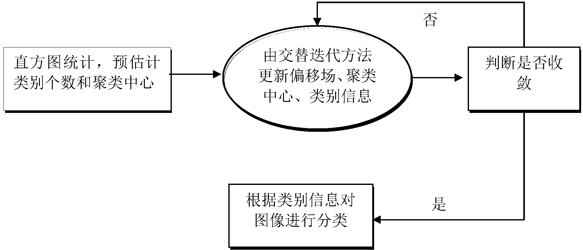
[0063] Step 1, firstly study the number of categories of breast MRI images, and increase the constraint of the smoothness of the offset field, and then combine the two to construct a coupling framework for the classification of breast tissue MRI images and offset field correction. The flow chart of this step like figure 1 shown:

[0064] Step 1.1, analyze the grayscale distribution of breast MRI to determine the number of categories of breast MRI images;

[0065] First, according to the gray distribution characterist...


PUM


Abstract

The invention discloses a breast tumor partition method. The breast tumor partition method includes the steps of building a coupled framework of classifications and biased field correcting of breast tissue nuclear magnetic resonance images, enhancing the breast areas and the peripheral areas, and partitioning the breast tumor images in cooperation with the shape prior. By means of the breast tumor partition method, biased field information is fused into classification models, the biased field information and the classification models are combined to be the unified framework, and the classifications and a corrected biased field of the breast tumor nuclear magnetic resonance images are solved with the fast energy minimization method at the same time, and use information of each other in the model evolution process for finally achieving accurate solving of the classifications and the corrected biased field; the shapes of blood vessels and tumors are analyzed, differences of the blood vessels and the tumors in shape are caught, the shape prior is built through parameters such as the characteristic values and the characteristic vectors, and the level set driving force based on the shapes is built and combined with a level set method based on local information, so that a level set overcomes the interference of a tubular structure during evolution and only captures the breast tumor area.

Description

technical field [0001] The invention belongs to the technical field of image segmentation, in particular to a method for extracting and segmenting breast tumor tissue based on nuclear magnetic resonance images. Background technique [0002] Breast cancer is the number one common malignancy in women, killing more than 10,000 women worldwide each year from the disease. In 2010, there were nearly 207,090 new breast cancer patients in the United States, and by 2011 this statistic had increased to 230,480 cases, accounting for 30% of new female malignant tumors, ranking first in the incidence of female malignant tumors. . In my country, statistics from big cities such as Beijing, Shanghai, and Tianjin show that breast cancer is also the most common malignant tumor among women in my country, and the incidence is increasing year by year. The symptoms of breast cancer are varied, common ones are: breast lumps, breast pain, nipple discharge, erosion or skin depression, swollen axil...

Claims


Application Information

Patent Timeline
Patent Type & Authority Applications(China)
IPC IPC(8): G06T7/00A61B5/055
CPCG06T7/11G06T2207/30068G06T2207/30096
Inventor 顾升华詹天明郑钰辉陈允杰罗君
Owner SHENZHEN BASDA MEDICAL APP
Why Eureka
Try Eureka