Separation method of liver cancer stem cells

A technology for liver cancer stem cells and liver cancer cells is applied in the field of cell separation, which can solve the problems of lack of specific marker identification and separation of LCSCs, and achieve the effects of good application prospects, improved success rate and simple operation.

Inactive Publication Date: 2015-04-22
YANGTZE NORMAL UNIVERSITY
View PDF0 Cites 2 Cited by
  • Summary
  • Abstract
  • Description
  • Claims
  • Application Information

AI Technical Summary

Benefits of technology

This patented technology allows researchers to use specific proteins called Fluorescence Antigens or FLU to tag targeted liverscancer cells together. These targets include labeled monoclonal antibodies like FCGR3 and LF4 linked through an oligonucleotide linkage between them. By comparing these labels from different types of liverycells they found differences in their ability to form colonies within hepatocytes. They also discovered how certain parts of those marked areas could be associated with other important tissue structures such as blood vessels, connective tissues, muscles, bone marrow stromal elements, etc., which may help predict treatment responses based on patient outcome data collected during clinic trials.

Problems solved by technology

The technical problem addressed by this patented technology relates to finding and distinguishing between different types of cancers called hepatocellular carcinoma or cholangiocytic disease caused due to gene mutations such as KRAS, PTEN loss, TP53 alteration, EGFR amplification etc., particularly with regards to developing effective treatments against them.

Method used


Image

  • Separation method of liver cancer stem cells
  • Separation method of liver cancer stem cells

Examples


Embodiment 1

[0027] Example 1. Isolation of liver cancer stem cells

[0028] To isolate liver cancer stem cells, follow the steps below:

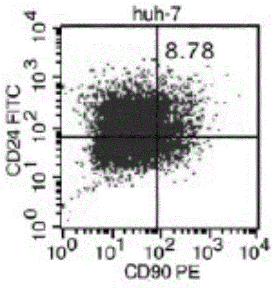
[0029] 1) Liver cancer cell line Huh-7 was cultured with 10% fetal bovine serum DMEM containing 100u / mL penicillin and streptomycin and high glucose at 37°C, 90% relative humidity, 5% CO 2 cultured in an incubator.

[0030] 2) When the cell coverage rate reaches 90%, the hepatoma cell culture solution obtained in step 1 is discarded, washed with PBS, digested with 0.25% trypsin until the cells fall off, the digestion is terminated, and passaged.

[0031] Cell subculture is performed as follows:

[0032] When it is observed that the confluence of cells in the culture flask is 90% (cell growth is in the logarithmic phase), the cells after successful thawing and resurrection are separated and cultured.

[0033] ① Discard the old culture medium and wash 1-2 times with PBS solution (without calcium and magnesium ions).

[0034] ② Add 1ml of digestion sol...

Embodiment 2

[0041] Example 2. Detecting spheroidizing ability of 4 kinds of liver cancer cell subpopulations

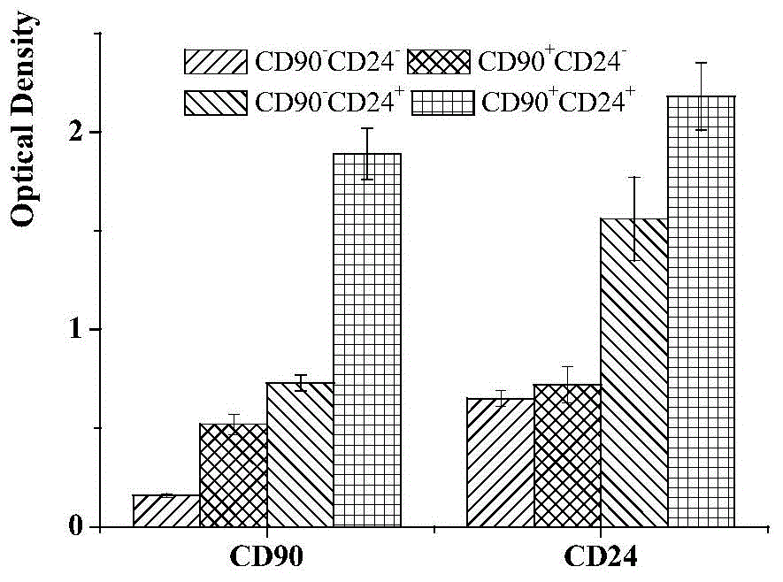
[0042]On the ultra-low adhesion culture plate, the four subgroups of liver cancer cells CD90+CD24+, CD90+CD24-, CD90-CD24+, CD90-CD24- were inoculated into DMEM / F12 medium without serum (containing 20 ng / mL bFGF (basic fibroblast growth factor, cationic polypeptide composed of 155 amino acids), 20ng / mL EGF (oligopeptide-1, active polypeptide composed of 53 amino groups), 20ng / mL LIF (leukemia inhibitory factor) , 2% B27 (cell culture additive), 100u / mL of penicillin and 100u / mL of streptomycin), observe the number of spheres of cells in 7 days, 14 days, 21 days and 28 days, and carry out the selected cells After culture and expansion, the cells were collected at the 3rd, 5th, 7th, 10th, and 13th passages, and the ability to form spheres was repeatedly verified three times. Using t statistical analysis, it was found that CD90+CD24+ liver cancer cells were significantly different f...

Embodiment 3

[0043] Example 3. Detection of clonal formation rate of 4 kinds of liver cancer cell subpopulations

[0044] The 4 kinds of liver cancer cells were inoculated in 6-well plates with 1000 cells per well, cultured in low-serum culture medium made of 5% FBS (fetal bovine serum) and double antibodies, the medium was changed every 2 days, and terminated after 3 weeks Culture, discard the supernatant, wash twice with PBS, fix with methanol, add Giemsa stain reagent 1mL / well, stain for 30min, then slowly wash away the staining solution with running water, and air dry. Finally, count the colonies with more than 50 cells under an ordinary light microscope, and calculate the colony formation rate (PE, PE=number of colonies / number of seeded cells×100%), CD90 + CD24 + The colony formation rate of liver cancer cells was (53.31±0.85)%, much higher than that of CD90 + CD24 - , CD90 - CD24 + , CD90 - CD24 - , with significant difference (P<0.01, P<0.001, P<0.001).


PUM


Abstract

The invention relates to a separation method of liver cancer stem cells. The separation method comprises the following steps: (1) culturing liver cancer cells by virtue of a DMEM high-glucose culture medium; (2) carrying out digestion passage on liver cancer cell culture fluid obtained in the step (1); and (3) marking passage cells being in a logarithmic phase during the digestion passage in the step (2) by virtue of flow sorting fluorescent antibodies CD90 and CD24, sorting by virtue of a flow cytometer, so as to obtain the separated liver cancer stem cells which are CD90+CD24+liver cancer cell subsets marked with CD90 and CD24. According to the separation method, two markers are utilized for carrying out joint detection, so that the separation and authentication successful rates of the liver cancer stem cells are increased; the operation is rapid, and the liver cancer stem cells can be rapidly and efficiently separated; the separation method is a new method for separating the liver cancer stem cells and has a good application prospect.

Description


Claims


Application Information

Patent Timeline
Owner YANGTZE NORMAL UNIVERSITY
Why Eureka
Try Eureka