Removal method and system for bone tissue in 3D CT (Three Dimensional Computed Tomography) image

A technology in 3DCT and images, applied in image enhancement, image analysis, image data processing, etc., can solve problems such as extraction performance impact

Inactive Publication Date: 2015-05-13
XIAN HWATECH MEDICAL INFORMATION TECH
View PDF4 Cites 17 Cited by
  • Summary
  • Abstract
  • Description
  • Claims
  • Application Information

AI Technical Summary

Problems solved by technology

Statistical shape methods can effectively suppress the influence of artifacts and noise generated in the imaging process, but the extraction performance is affected by the accuracy of the average shape learned. At the same time, for bone tissue data, the shape labeling and training process It requires a lot of manpower and time

Method used


Image

  • Removal method and system for bone tissue in 3D CT (Three Dimensional Computed Tomography) image
  • Removal method and system for bone tissue in 3D CT (Three Dimensional Computed Tomography) image
  • Removal method and system for bone tissue in 3D CT (Three Dimensional Computed Tomography) image

Examples


Embodiment 1

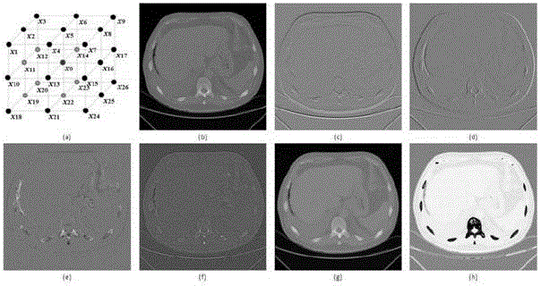
[0148] In this embodiment, test cases Pat_4, Pat_5, and Pat_2 are tested for the supervoxel segmentation process, and the results of supervoxel segmentation for each case are as follows Figure 7 shown. After supervoxel segmentation, some supervoxel segmentation results are as follows Figure 6 shown. Depend on Figure 7 and Figure 6 It can be seen that with the threshold T=[t l ,t h ], the number of supervoxels decreases sharply, and at the same time, the segmentation of supervoxels takes less and less time.

Embodiment 2

[0150] In this example, bone extraction and removal tests were performed on 7 groups of cases. In this test example, some supervoxels in cases Pat_2, Pat_3 and Pat_4 are marked for random forest classifier training, and the specific supervoxel marking information is shown in Table 1. Bone extraction and removal results such as Figure 8 shown. The bone tissue extraction and removal results of Pat_5, Pat_6, Pat_7 and Pat_8 are as follows: Figure 9 shown. Depend on Figure 8 , Figure 9 It can be seen that the system of the present invention can obtain satisfactory extraction and removal results without any prior knowledge of the shape of bone tissue.


PUM


Abstract

The invention discloses a removal method and system for a bone tissue in a 3D CT (Three Dimensional Computed Tomography) image. The method comprises the following steps: reading a 3D CT image data sequence and dividing the sequence by using a clustering method; describing each marked supervoxel from supervoxel level, pixel level and supervoxel neighborhood relation level respectively to obtain a characteristic vector for describing each supervoxel; constructing a supervoxel training set, training a classifier by using the characteristic vector corresponding to the supervoxel, then classifying the supervoxel by using the trained classifier and marking the category; if a certain supervoxel is marked as the bone tissue according to a supervoxel mark value, removing the supervoxel from the original CT data, and otherwise, remaining. The supervoxel obtained by dividing the 3D CT data is used as a basic operation unit, bone tissue templates on different parts are not required to be learnt and trained, and meanwhile the problem of inaccurate bone tissue extraction caused by mismatching of the templates is also avoided.

Description

technical field [0001] The invention belongs to the technical field of target retrieval and extraction, and in particular relates to a method and system for removing bone tissue in a 3D CT image. The invention is used for automatically extracting and removing bone tissue from 3D CT data collected by CT imaging equipment. Background technique [0002] Compared with X-ray images, CT (computed tomography) can provide doctors with complete and high-resolution 3D information about target organs, and it has become an important tool for clinical observation at present. With the development of digital clinical technology, an effective computer-aided diagnosis and decision-making system is of great significance to doctors' reasonable and rapid diagnosis and formulation of diagnosis and treatment plans. The extraction and segmentation of different organs is an important step in the accurate interpretation of CT images in many applications. A large number of segmentation and classifi...

Claims


Application Information

Patent Timeline
Patent Type & Authority Applications(China)
IPC IPC(8): G06T7/00
CPCG06T7/0012G06T2207/10012G06T2207/10081G06T2207/20081G06T2207/30008
Inventor 赵志强申田李云峰张孝林
Owner XIAN HWATECH MEDICAL INFORMATION TECH
Why Eureka
Try Eureka