Automatic extraction method of three-dimensional breast full-volume image regions of interest

A region of interest, three-dimensional ultrasound technology, applied in the field of image processing, can solve problems such as dependence and time-consuming

Active Publication Date: 2015-05-27
FUDAN UNIV
View PDF5 Cites 49 Cited by
  • Summary
  • Abstract
  • Description
  • Claims
  • Application Information

AI Technical Summary

Problems solved by technology

This manual calibration method is very time-...

Method used


Image

  • Automatic extraction method of three-dimensional breast full-volume image regions of interest
  • Automatic extraction method of three-dimensional breast full-volume image regions of interest
  • Automatic extraction method of three-dimensional breast full-volume image regions of interest

Examples


Embodiment Construction

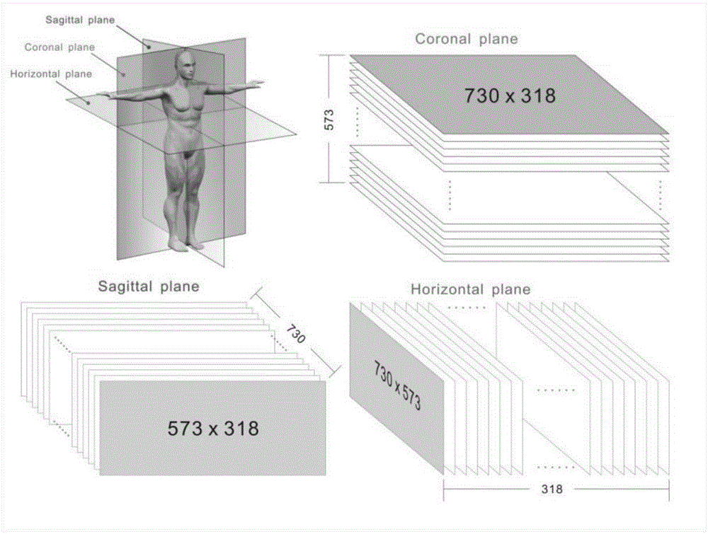
[0098] The method for automatically extracting the region of interest in the three-dimensional ultrasonic breast volume imaging (ABVS) proposed by the present invention is tested. ABVS image taken from ACUSON S2000 of Siemens AG TM Ultrasound instrument. The system is equipped with a broadband linear probe (14L5BV), which can obtain breast volume images of 15.4 cm×16.8 cm×(2~6) cm. A total of 15 ABVS images were collected in this experiment, each with 98-294 coronal images, 820 transverse images, and 750 sagittal images.

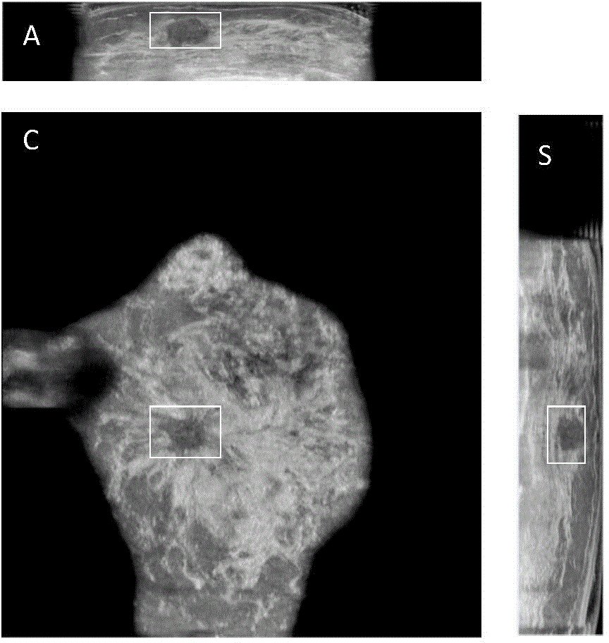
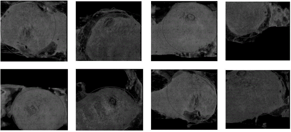
[0099] First, the original ABVS image is reconstructed, and the three sections (transverse, sagittal, and coronal) of the reconstructed image are as follows: figure 2 Shown, where the approximate outline of the breast can be seen on the coronal image. Depend on figure 2 It can be seen that the breast contour is generally close to an ellipse, so the Hough transform is used to find the ellipse representing the breast on the coronal image, such as image 3...


PUM


Abstract

The invention belongs to the field of image processing, and particularly relates to an automatic extraction method of regions of interest in three-dimensional breast full-volume images (ABVS). The method comprises the following steps: processing the continuous cross section two-dimensional images in three-dimensional ABVS images by using a maximum direction-based phase information method to obtain the candidate regions of interest on each cross section image; removing the unrelated regions according to the prior knowledge such as the continuity and position characteristic of breast tumor on the two-dimensional cross section images; obtaining the shape and texture features of the residual suspected tumor regions, inputting the shape and texture shapes to a two-valued logistic regression classifier to obtain the probability of each region becoming tumor and selecting the region with the maximum probability as the tumor region; obtaining the minimum ellipsoid comprising the region of interest according to the selected region to serve as the region of interest. The automatic extraction method provided by the invention can be used for realizing the automatic extraction of tumor regions of interest in the three-dimensional ABVS images, obtaining the correct positions of tumor, decreasing the workload of the manual operation and providing important reference to further tumor detection.

Description

technical field [0001] The invention belongs to the technical field of image processing, in particular to an automatic extraction method for a region of interest in a three-dimensional ultrasonic breast full-volume image. Background technique [0002] Ultrasound imaging has important clinical applications due to its advantages of non-invasive, real-time, strong repeatability, and low cost. Compared with the traditional handheld two-dimensional ultrasound imaging, ABVS has a new imaging mode, which can standardize and automatically scan the breast, digitally process the image, and avoid individual differences among users; ABVS can scan the whole breast, compared with conventional ultrasound, The reconstructed coronal section is added, which can provide more information than two-dimensional images, and has good repeatability. [0003] Since the volume of the tumor is relatively small compared to the entire ABVS image, the accuracy of direct tumor segmentation is low. Therefo...

Claims


Application Information

Patent Timeline
IPC IPC(8): G06T7/00G06T7/60A61B8/00
CPCA61B8/00A61B8/0825A61B8/085A61B8/52G06T7/0012G06T2207/10136G06T2207/30068
Inventor 汪源源王欣郭翌余锦华
Owner FUDAN UNIV
Why Eureka
Try Eureka