Automatic segmenting method for lesion tissue in lung CT image

A technology of CT imaging and lungs, applied in image analysis, image data processing, instruments, etc., can solve problems such as inaccuracy

Active Publication Date: 2015-06-17
INST OF AUTOMATION CHINESE ACAD OF SCI
View PDF4 Cites 12 Cited by
  • Summary
  • Abstract
  • Description
  • Claims
  • Application Information

AI Technical Summary

Problems solved by technology

The existing segmentation algorithms for lung lesion tissue based on region gro...

Method used


Image

  • Automatic segmenting method for lesion tissue in lung CT image
  • Automatic segmenting method for lesion tissue in lung CT image
  • Automatic segmenting method for lesion tissue in lung CT image

Examples


Embodiment Construction
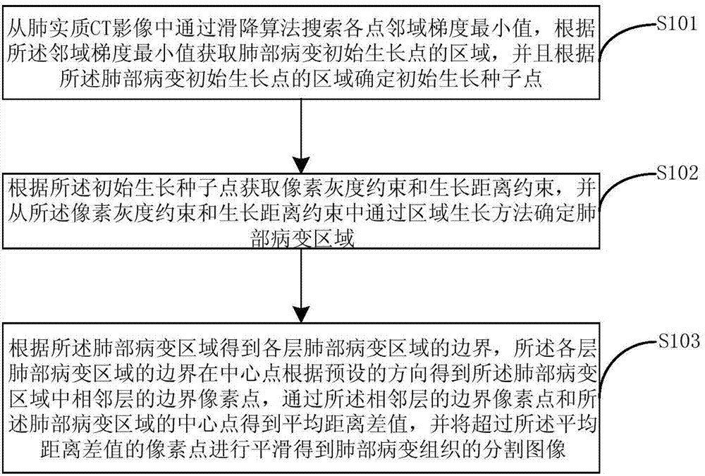
[0014] The general idea of ​​the present invention is to obtain the pixel grayscale constraint and the growth distance constraint according to the initial growth seed point, and determine the lung lesion area through the region growth method from the pixel grayscale constraint and the growth distance constraint, and analyze the lung lesion area. Smoothing is performed to accurately obtain segmented images of lung diseased tissue.

[0015] The following will describe in detail the automatic segmentation method of lung CT image lesion tissue with reference to the accompanying drawings.

[0016] figure 1 The flow chart of the automatic segmentation method for lung CT image lesion tissue provided by the embodiment of the present invention.

[0017] refer to figure 1 , in step S101, from the CT image of the lung parenchyma, the minimum value of the neighborhood gradient of each point is searched by the toboggan algorithm, and the area of ​​the initial growth point of the lung les...


PUM


Abstract

The invention provides an automatic segmenting method for lesion tissue in a lung CT image. The automatic segmenting method comprises the steps that the lung parenchyma CT image is searched for the minimum neighborhood gradient value of all points through a toboggan algorithm, the area of the initial growth point of the lung lesion is obtained according to the minimum neighborhood gradient value, and the initial growth seed point is determined according to the area of the initial growth point of the lung lesion; pixel grey level constraints and growth distance constraints are obtained according to the initial growth seed point, a lung lesion area is determined from the pixel grey level constraints and the growth distance constraints through an area growth method; boundaries of all layers of the lung lesion area are obtained according to the lug lesion area, boundary pixel points of adjacent layers in the lung lesion area are obtained in the central points of the boundaries of all the layers of the lung lesion area in the preset direction, the average distance difference value is obtained according to the boundary pixel points of the adjacent layers and the central point of the lung lesion area, and the pixel points exceeding the average distance difference value are horizontally slide to obtain segmentation images of the lung lesion tissue.

Description

technical field [0001] The invention relates to image segmentation technology, in particular to an automatic segmentation method for lung CT image lesion tissue. Background technique [0002] The automatic segmentation of lung CT image lesions is an important issue in the field of computer-aided diagnosis. It can automatically acquire lung lesions and shorten the waiting time for clinical diagnosis. It has important application value in the field of computer-aided diagnosis. [0003] Lung CT imaging lesions can be classified into isolated types, vascular adhesion types, and pleural adhesion types according to the location of the lesions; according to the types of lesions, they can be divided into ground-glass opacities, pulmonary nodules, and lung tumors. Among them, the ground-glass opacity type is the most difficult to segment in the lung lesions. Existing segmentation algorithms for lung lesions based on region growth all require manual acquisition of initial growth seed...

Claims


Application Information

Patent Timeline
IPC IPC(8): G06T7/00
Inventor 田捷宋江典杨彩云杨凤
Owner INST OF AUTOMATION CHINESE ACAD OF SCI
Why Eureka
Try Eureka