
Breast ultrasound scanning device

A technology for ultrasound scanning, breast, applied in the field of medical imaging, can solve problems such as poor image quality

Inactive Publication Date: 2015-12-23
王士平
View PDF13 Cites 8 Cited by
  • Summary
  • Abstract
  • Description
  • Claims
  • Application Information

AI Technical Summary

Problems solved by technology

This presents two major disadvantages: (1) poor image quality; and (2) limited range of breast sizes that can be scanned

Method used


Image

  • Breast ultrasound scanning device
  • Breast ultrasound scanning device
  • Breast ultrasound scanning device

Examples


Embodiment Construction

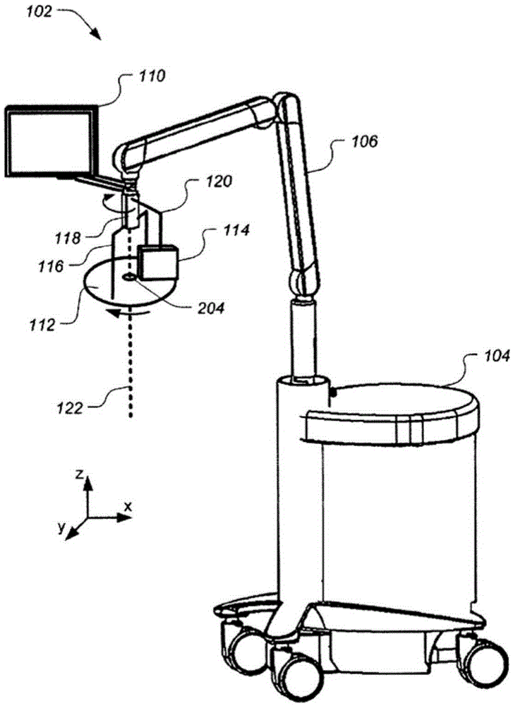
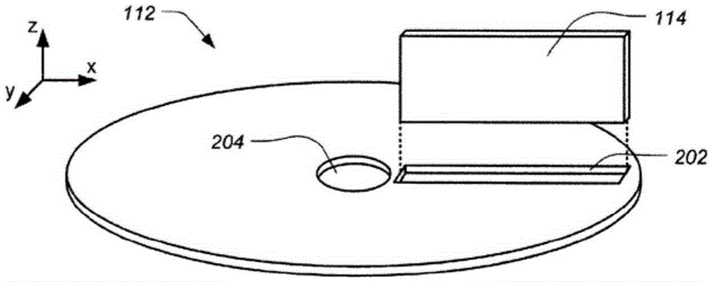
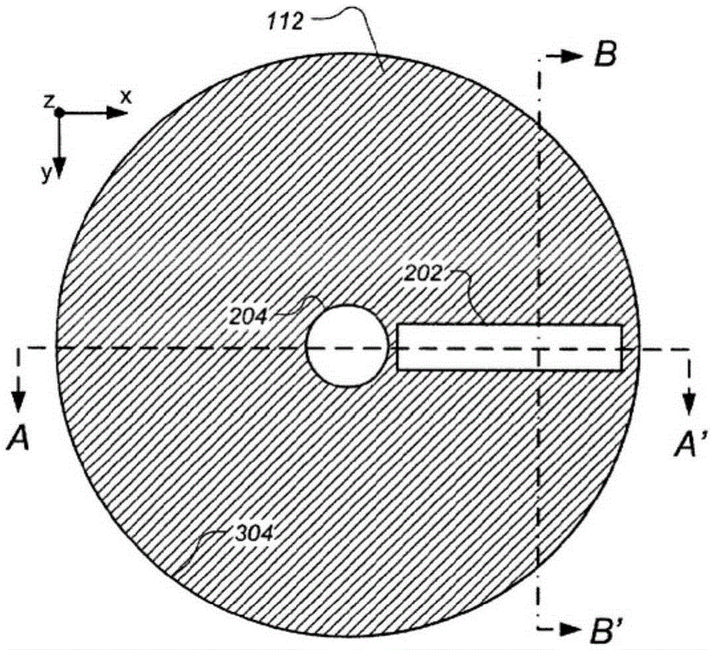
[0042] figure 1 Illustrated is a perspective view of a full field breast ultrasound (FFBU) scanning apparatus 102 comprising a frame 104 which may contain an ultrasound processor, a movable support arm 106 and a monitor 110 attached to the support arm 106 in accordance with a preferred embodiment. The FFBU scanning device 102 also includes a substantially planar radial scan template 112 and an ultrasound transducer 114 . Radial scan template 112 is configured to compress the breast of a patient (eg, a supine patient) chest-to-chest while rotating about axis 122 , preferably centered on nipple aperture 204 . The ultrasound transducer 114 rotates with the radial scanning plane template 112 and scans the breast through slot-shaped radially extending openings in the template. For reference purposes here, the +z direction refers to the outward direction away from the patient's chest, the x-axis refers to the side-to-side direction relative to a supine patient, and the y-axis refe...


PUM


Abstract

An apparatus and a method are disclosed for obtaining ultrasound images of a patient's breast that is chestwardly compressed with a template that is essentially planar and rotates relative to the breast while one or more ultrasound transducers moving with the template take 2D images of the breast through one or more respective radially oriented slots in the template, preferably through a membrane that is porous to a gel. The 2D images are processed into slice images representing breast slices of desired thicknesses and orientation that are displayed alone or with some of the 2D images, preferably pairs of orthogonally disposed 2D images.

Description

[0001] Cross References to Related Applications [0002] This application claims priority to US Provisional Application 61 / 769,913, filed February 27, 2013. This application is a continuation-in-part of U.S. Application Serial No. 13 / 296,023 filed November 14, 2011 (published as US2012 / 0089026A1 and scheduled to be issued as US Patent 8,579,819 on November 12, 2013), U.S. Application Serial No. 13 / 296,023 is a continuation of US Application Serial No. 11 / 513,481 filed August 30, 2006 and claiming the benefit of US Provisional Application No. 60 / 713,282, filed September 1, 2005. The entire contents of all of the above applications are hereby incorporated by reference. The entire contents of the patent applications and patents and other publications cited below are also incorporated herein by reference. technical field [0003] This patent specification relates to medical imaging. More specifically, this patent specification relates to ultrasound imaging of the breast using c...

Claims


Application Information

Patent Timeline
Patent Type & Authority Applications(China)
IPC IPC(8): A61B8/00
CPCA61B8/0825A61B8/403A61B8/4209A61B8/4461A61B8/4477A61B8/463A61B8/466A61B8/483A61B8/4218A61B8/085A61B8/4405A61B8/523
Inventor 王士平
Owner 王士平
Learn More