Ultrasonic imaging method and device and ultrasonic equipment comprising ultrasonic imaging device

An ultrasonic image, the same technology, applied in ultrasonic/sonic/infrasonic diagnosis, ultrasonic diagnosis, infrasonic diagnosis, etc., can solve the problems of high missed diagnosis rate, difficulty in detecting fetal ear position, and displaying fetus at the same time, so as to avoid missed diagnosis.

Active Publication Date: 2016-03-30
SONOSCAPE MEDICAL CORP
View PDF3 Cites 8 Cited by
  • Summary
  • Abstract
  • Description
  • Claims
  • Application Information

AI Technical Summary

Problems solved by technology

[0004] The display of fetal ear is affected by amniotic fluid volume, fetal position and fetal umbilical cord, etc., and two-dimensional ultrasound cannot simultaneously display the fetal ear and eye in one section, making it difficult to detect abnormal fetal ear position, and the rate of missed diagnosis is high.

Method used


Image

  • Ultrasonic imaging method and device and ultrasonic equipment comprising ultrasonic imaging device
  • Ultrasonic imaging method and device and ultrasonic equipment comprising ultrasonic imaging device
  • Ultrasonic imaging method and device and ultrasonic equipment comprising ultrasonic imaging device

Examples


Embodiment 1

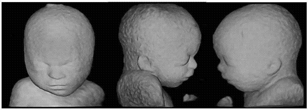
[0033] The auricle is mostly composed of cartilage, and is generally bilaterally symmetrical. It is shaped like a shell and forms an angle of about 30° with the skull. At the 20th week of embryonic development, the shape of the auricle is similar to an adult. The position of the auricle was initially low when it was formed, which is equivalent to the upper part of the future neck. With the development of the mandible, the position of the auricle gradually rises to about the height of the flat eyes on both sides of the head. If this developmental obstacle occurs, you can Forms lower ears.

[0034] Lower ears can be found in Alpert syndrome, DiGeorge syndrome, Pierre syndrome, trisomy 21, etc., and are common in inherited middle ear malformations, inner ear malformations caused by multiple chromosomal aberrations, and loss of inner ear hair cells. Two-dimensional ultrasound cannot display the auricle and eye of the fetus at the same time on one slice, so it is very easy to miss the...

Embodiment 2

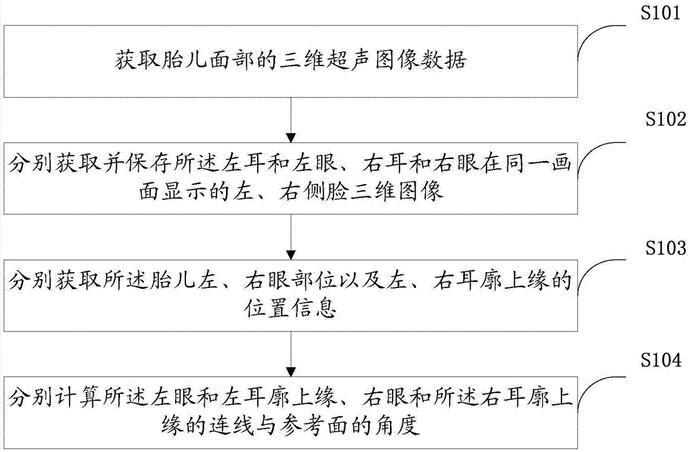
[0049] In some embodiments, in the step S102, before separately acquiring and saving the left and right face three-dimensional images displayed on the same screen by the left ear and left eye, right ear and right eye, it may also include correcting the fetus Steps for facial image to horizontal position.

[0050] After acquiring the 3D ultrasound image of the fetal face, due to the position of the fetus, the 3D image of the fetus displayed on the screen may be tilted. When the fetus is tilted, it may cause trouble in the subsequent steps or cause inaccurate measurement. Therefore, the fetal face can be rotated to the horizontal level before obtaining the eye position information to overcome the above-mentioned shortcomings.

[0051] There are many ways to correct the fetal facial image to the level. For example, to find the reference object, the line between the two eyes can be used as the reference object, and the line between the two eyes (usually the center of the two eyes) and ...

Embodiment 3

[0053] In some embodiments, in the step S102, before separately acquiring and saving the left and right face three-dimensional images displayed on the same screen by the left ear and left eye, right ear and right eye, it may also include acquiring a fetal face image The middle of the steps.

[0054] After acquiring the three-dimensional ultrasound image of the fetus’s face, if it can be ensured that the fetus’s face is in the median plane, a uniform angle can be set to control the fetal facial image to rotate to the left and right, so as to obtain the left ear and Side images of the left and right faces of the left eye, right eye and right ear.


PUM


Abstract

The invention provides an ultrasonic imaging method. The method comprises the steps that three-dimensional ultrasound image data of the face of a fetus are obtained; three-dimensional images, displayed on the same picture, of the left eye and ear and right ear and eye of the left side and the right side of the face are obtained respectively; the position information of the left and right eyes and the position information of the upper edges of the left and right auricles are obtained respectively; the angle between the connecting line of the left eye and the upper edge of the left auricle and the reference face and the angle between the connecting line of the right eye and the upper edge of the right auricle and the reference face are calculated respectively. The invention further provides an ultrasonic imaging device and ultrasonic equipment comprising the same. The low-set ear condition of the fetus can be rapidly diagnosed, and missed diagnosis is avoided.

Description

Technical field [0001] The present invention relates to the technical field of ultrasound imaging, in particular to an ultrasound imaging method, device and ultrasound equipment. Background technique [0002] The auricle is mostly composed of cartilage, and is generally bilaterally symmetrical. It is shaped like a shell and forms an angle of about 30° with the skull. At the 20th week of embryonic development, the shape of the auricle is similar to an adult. The position of the auricle is low when it is first formed, which is equivalent to the upper part of the future neck. With the development of the mandible, the position of the auricle gradually rises to the height of the flat eyes on both sides of the head. If this developmental obstacle occurs, it can be Forms lower ears. [0003] Lower ears can be found in Alpert syndrome, DiGeorge syndrome, Pierre syndrome, 21-trisomy syndrome, etc., and are common in inherited middle ear malformations, inner ear malformations caused by mult...

Claims


Application Information

Patent Timeline
Patent Type & Authority Applications(China)
IPC IPC(8): A61B8/00
Inventor 李萍刘旭江许龙唐艳红党静
Owner SONOSCAPE MEDICAL CORP
Why Eureka
Try Eureka