Ultrasonic imaging method, device and ultrasonic equipment thereof
An ultrasound image and three-dimensional ultrasound technology, which is applied in ultrasound/sound wave/infrasonic wave diagnosis, sound wave diagnosis, infrasonic wave diagnosis, etc., can solve the problems of high missed diagnosis rate, difficulty in detecting fetal ear position, and displaying fetus at the same time, so as to avoid missed diagnosis Effect
- Summary
- Abstract
- Description
- Claims
- Application Information
AI Technical Summary
Problems solved by technology
Method used
Image
Examples
Embodiment 1
[0033] Most of the auricle is composed of cartilage, generally symmetrical on both sides, shaped like a shell, and forms an angle of about 30° with the skull. At the 20th week of embryonic development, the shape of the auricle is similar to that of an adult. The position of the auricle is low when it is first formed, which is equivalent to the upper part of the future neck. With the development of the mandible, the position of the auricle gradually rises to the height of the eye level on both sides of the head. Form low ears.
[0034] Low-set ears can be seen in Alpert syndrome, DiGeorge syndrome, Pierre syndrome, trisomy 21, etc., and are common in diseases such as hereditary middle ear deformities, inner ear deformities caused by various chromosomal aberrations, and loss of inner ear hair cells. Two-dimensional ultrasound cannot simultaneously display the auricle and eyes of the fetus in one section, so it is very easy to miss the diagnosis clinically.
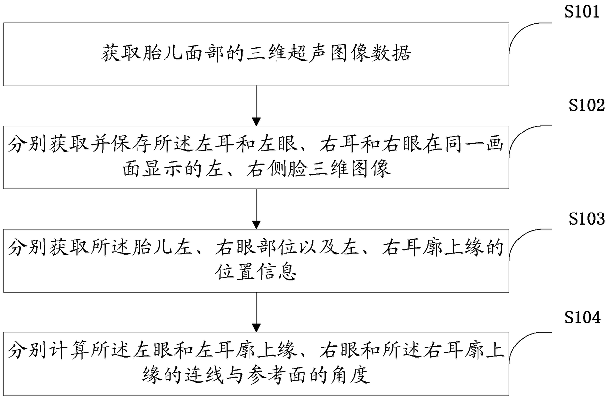
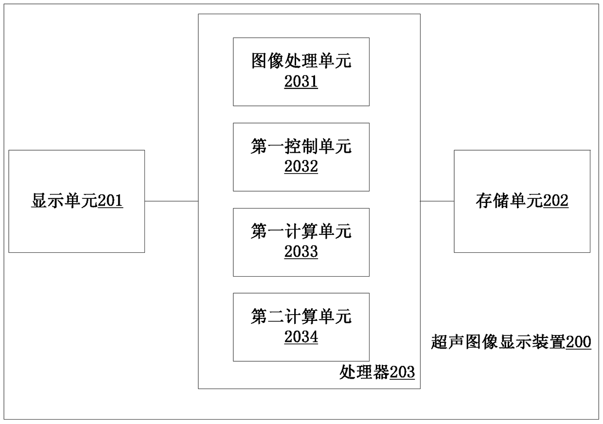
[0035] Such as fig...
Embodiment 2
[0049] In some embodiments, in the step S102, before obtaining and saving the left ear and the left eye, the right ear and the right eye, and the left and right face three-dimensional images displayed on the same screen, it may also include correcting the fetus Steps for face image to horizontal position.
[0050]After obtaining the 3D ultrasound image of the fetal face, due to the position of the fetus, the 3D image of the fetus displayed on the screen may be tilted. Judging in the tilted state of the fetus may cause trouble in the subsequent steps or cause inaccurate measurements. Therefore, the fetal face can be rotated to the horizontal before obtaining the eye position information, so as to overcome the above defects.
[0051] There are many ways to correct the fetal facial image to the level, for example: the method of finding the reference object, the connection line between the two eyes can be used as the reference object, and the connection line between the two eyes (...
Embodiment 3
[0053] In some embodiments, in the step S102, before obtaining and storing the left ear and left eye, right ear and right eye three-dimensional images of the left and right faces displayed on the same screen, it may also include obtaining fetal facial images The steps on the median side.
[0054] After obtaining the three-dimensional ultrasonic image of the fetal face, if it can be ensured that the face of the fetus is located in the median plane, a uniform angle can be set to control the rotation of the fetal facial image to the left and right, so as to obtain the images including the left ear and the right ear respectively. Left and right side profile images of the left eye and the right eye and ear.
PUM
 Abstract
Description
Claims
Application Information
 - R&D
- Intellectual Property
- Life Sciences
- Materials
- Tech Scout
- Unparalleled Data Quality
- Higher Quality Content
- 60% Fewer Hallucinations
Browse by: Latest US Patents, China's latest patents, Technical Efficacy Thesaurus, Application Domain, Technology Topic, Popular Technical Reports.
© 2025 PatSnap. All rights reserved.Legal|Privacy policy|Modern Slavery Act Transparency Statement|Sitemap|About US| Contact US: help@patsnap.com


