
Method and apparatus for processing medical image

A medical imaging and imaging technology, applied in the field of medical imaging, can solve the problems of not being able to completely define adjacent areas, not accurately conforming to anatomical structures, and being inaccurate

Inactive Publication Date: 2016-08-03
BEIJING QED TECHN CO LTD
View PDF7 Cites 6 Cited by
  • Summary
  • Abstract
  • Description
  • Claims
  • Application Information

AI Technical Summary

Problems solved by technology

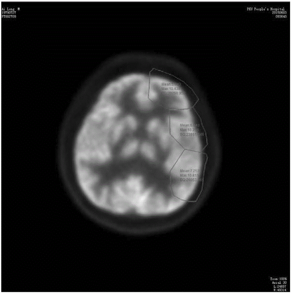
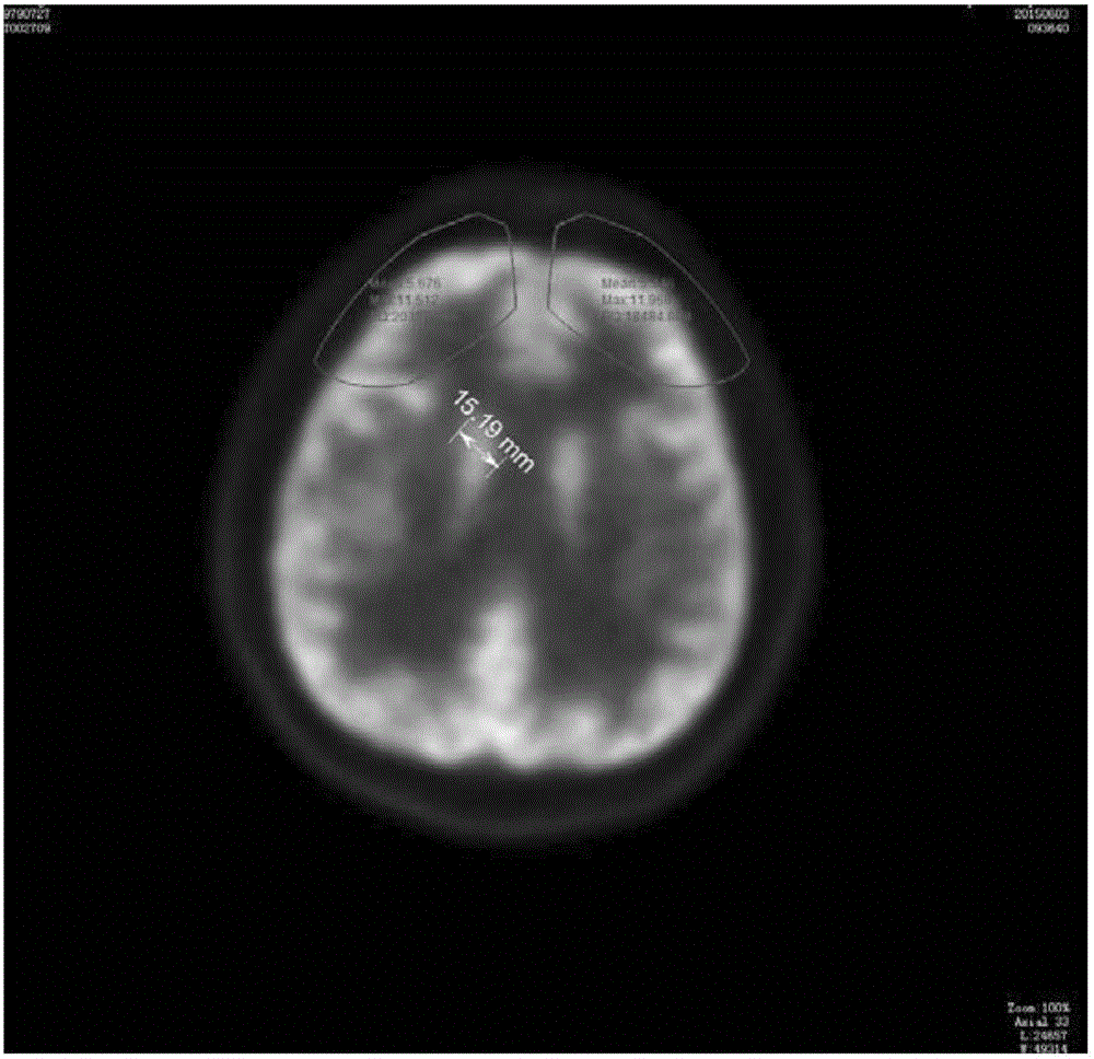
However, in the related prior art for drawing ROI, the shape of the drawn area can only roughly delineate the functional area to be drawn, and drawing shapes such as a circle, ellipse or rectangle cannot precisely conform to the anatomical structures, and cannot perfectly define adjacent areas, such as figure 2 shown
Since such ROI does not reflect the actual internal structure of related organs well, and the values ​​obtained based on this ROI area are incomplete or inaccurate, resulting in inaccurate subsequent processing and analysis results of medical images, which unfavorably limits The range of current medical imaging applications

Method used


Image

  • Method and apparatus for processing medical image
  • Method and apparatus for processing medical image
  • Method and apparatus for processing medical image

Examples


Embodiment Construction

[0064] The present invention will be further described through specific embodiments below in conjunction with the accompanying drawings. It should be understood that the following content is only for explaining and illustrating the present invention, and will not limit the present invention in any respect.

[0065] In this document, unless specifically stated otherwise, the terms "upper", "lower", "left", "right", "front", "rear", "inner", "outer", "transverse", "longitudinal" , "middle", "sideways" and so on are descriptions relative to the directions shown on the pages of the accompanying drawings.

[0066] In this article, the terms "first", "second", etc. are used only to distinguish different parts or steps, to indicate that these parts or steps are independent of each other, but not to explain that there is an important relationship between these parts or steps. Restrictions on gender, order, position, etc.

[0067] In one aspect, the invention relates to mapping a reg...


PUM


Abstract

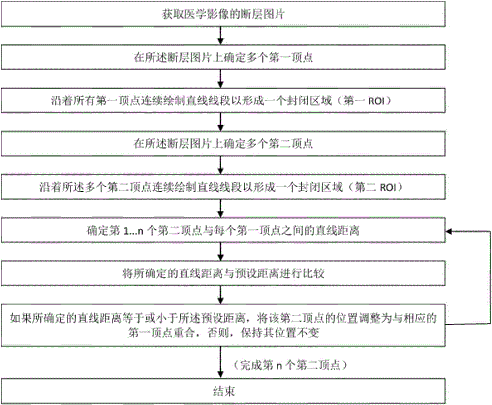
The invention relates to a method for processing a medical image. The method comprises the following advantages: obtaining a tomogram of the medical image; drafting a first ROI and a second ROI on the tomogram; and performing overlapping processing on the first ROI and the second ROI so as to enable the portions, which are corresponding to a continuous tissue structure, on first ROI and the second ROI to be connected together. The invention also relates to an apparatus for processing a medical image.

Description

technical field [0001] The invention relates to medical image technology, in particular to a device and method for processing medical images. Background technique [0002] The development of modern medical imaging has provided a large amount of practical anatomical and functional imaging data for clinical practice, and has become an indispensable part of modern medical care. At present, the application of medical imaging runs through the entire clinical work. It is not only widely used in disease diagnosis, but also plays an important role in planning design, program implementation and efficacy evaluation of surgical operations and radiotherapy. [0003] Medical imaging refers to the technology and processing process of obtaining internal tissue images of the human body or a certain part of the human body in a non-invasive manner for medical treatment or medical research. Since the invention of X-rays, the development of medical imaging technology has gone through three sta...

Claims


Application Information

Patent Timeline
IPC IPC(8): G06T7/00
CPCG06T2207/10084G06T2207/30016
Inventor 李刚
Owner BEIJING QED TECHN CO LTD
Learn More