3D printing preparation method of small-aperture biological artificial blood vessel and artificial blood vessel

A bioartificial, 3D printing technology, applied in the field of implanted devices in medical devices, can solve the problems of cell death and high printing temperature, and achieve the effects of avoiding cell death, slowing down shaking, and improving printing quality

Inactive Publication Date: 2017-06-09
广州尤尼智康生物科技有限公司
View PDF2 Cites 12 Cited by
  • Summary
  • Abstract
  • Description
  • Claims
  • Application Information

AI Technical Summary

Problems solved by technology

[0005] This application provides a 3D printing preparation method of a small-diameter bioartificial blood vessel and an artificial blood vessel, which solves the problem that the printing temperature of the existing scaffold material is higher than the survival temperature of the cells in the process of printing small-diameter bioartificial blood vessels using 3D technology. technical problems of death

Method used


Image

  • 3D printing preparation method of small-aperture biological artificial blood vessel and artificial blood vessel
  • 3D printing preparation method of small-aperture biological artificial blood vessel and artificial blood vessel
  • 3D printing preparation method of small-aperture biological artificial blood vessel and artificial blood vessel

Examples


Embodiment Construction

[0022] The specific implementation manners of the present application will be described in further detail below in conjunction with the accompanying drawings.

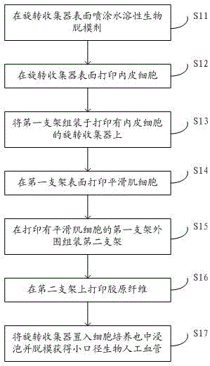
[0023] The 3D printing preparation method of the small-caliber bioartificial blood vessel proposed by this application, such as figure 1 shown, including the following steps:

[0024] Step S11: Spraying a water-soluble biological release agent on the surface of the rotating collector.

[0025] Such as figure 2 As shown, before printing the artificial blood vessel, spraying the water-soluble biological release agent 21 on the surface of the rotating collector 31 can solve the problem that the printed artificial blood vessel is not easy to release from the mold. Water-soluble bio-release agents such as water-soluble chitosan, water-soluble lactic acid, etc., do not affect biocompatibility.

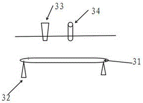
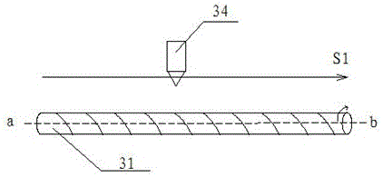
[0026] Such as image 3 As shown, the rotating collector 31 is the main carrier of the printed artificial blood vessel, which ...


PUM

PropertyMeasurementUnit
diameteraaaaaaaaaa
thicknessaaaaaaaaaa

Abstract

The invention discloses a 3D printing preparation method of a small-aperture biological artificial blood vessel and an artificial blood vessel to solve the technical problem that the printing temperature of a stent material is higher than the survival temperature of cells, thus causing cell death in a process of printing a small-aperture biological artificial blood vessel by a 3D printing technology. After endothelial cells are printed on the surface of a rotary collector, a first stent is assembled on the rotary collector with the printed endothelial cells, then smooth muscle cells are printed on the surface of the first stent, a second stent is assembled on the periphery of the first stent with the printed smooth muscle cells, and collagenous fiber is printed on the second stent; and finally, the rotary collector is soaked in a cell culture dish, and demoulding is performed to obtain a small-aperture biological artificial blood vessel. The first stent and the second stent are prepared in advance by a 3D printing method; and the pre-preparation way of the two stents can ensure that the cells are assembled, printed and seeded after the stent cooling, the layered uniform seeding of cells is guaranteed while the problem of cell death caused by the high printing temperature is avoided.

Description

technical field [0001] The invention belongs to the technical field of implanted devices in medical devices, and in particular relates to a 3D printing preparation method of a small-diameter bioartificial blood vessel and an artificial blood vessel. Background technique [0002] At present, most of the artificial blood vessels used clinically are non-degradable polymer materials such as polyester products and polytetrafluoroethylene. For the replacement of large blood vessels, these artificial blood vessels meet the clinical needs to a certain extent. However, for the replacement of small blood vessels with a diameter of less than 6 mm, the artificial blood vessels of this material can achieve a better patency effect in the early stage by coating the inner surface with anticoagulant or antithrombotic drugs, but the drugs are gradually released in the later stage of transplantation. After completion, it is easy to form a blood clot and eventually lead to the failure of the v...

Claims


Application Information

Patent Timeline
Patent Type & Authority Applications(China)
IPC IPC(8): A61L27/50A61L27/38A61L27/24B33Y10/00B33Y80/00B33Y70/00
CPCA61L27/507A61L27/24A61L27/3808A61L27/3826B33Y10/00B33Y70/00B33Y80/00
Inventor 王红施敏超胡海龙
Owner 广州尤尼智康生物科技有限公司
Why Eureka
Try Eureka