Segmentation method of blood pool in end-diastolic images of cardiac functional magnetic resonance images

A technology of magnetic resonance imaging and end-diastole, which is applied in the field of medical imaging and can solve problems such as complexity and low efficiency

Active Publication Date: 2020-11-27
SHANGHAI UNITED IMAGING HEALTHCARE
View PDF4 Cites 0 Cited by
  • Summary
  • Abstract
  • Description
  • Claims
  • Application Information

AI Technical Summary

Problems solved by technology

[0003] Determination of end-diastole in 4D fMRI images is often manual and user-assisted semi-automatic methods that are cumbersome, inefficient, and subject to large inter-observer and intra-observer variability

Method used


Image

  • Segmentation method of blood pool in end-diastolic images of cardiac functional magnetic resonance images
  • Segmentation method of blood pool in end-diastolic images of cardiac functional magnetic resonance images
  • Segmentation method of blood pool in end-diastolic images of cardiac functional magnetic resonance images

Examples


Embodiment Construction

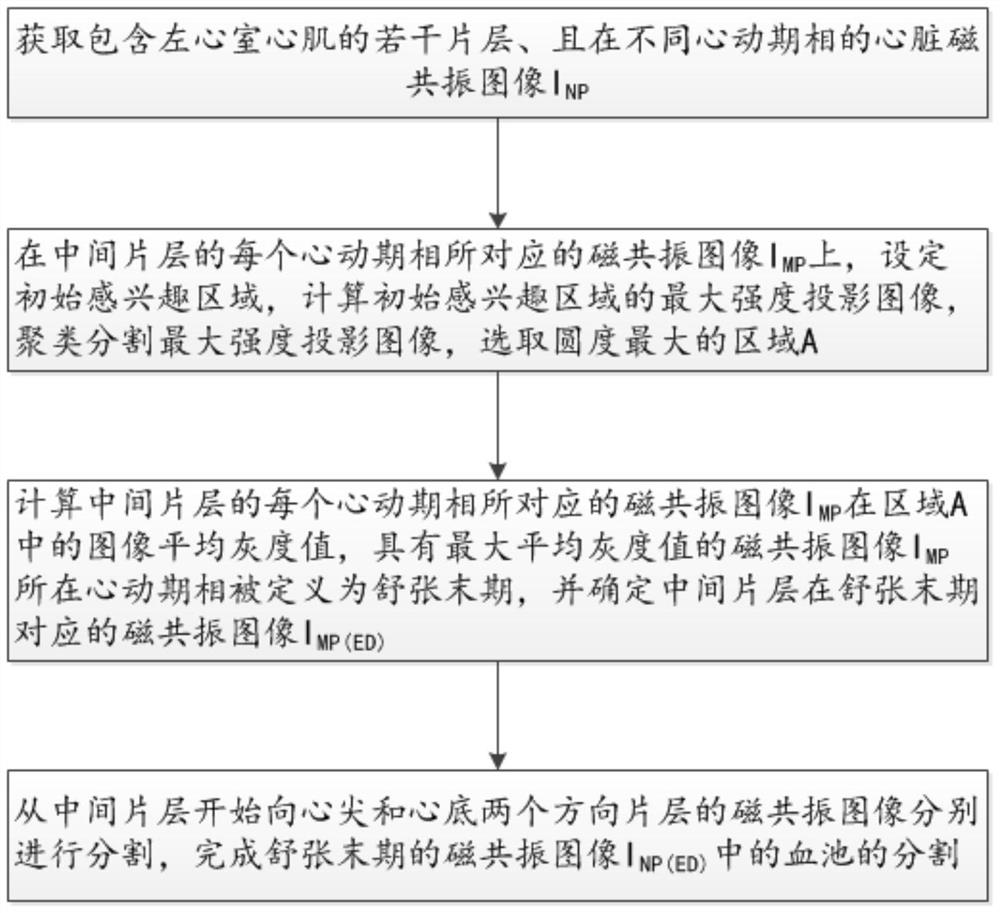
[0036] see figure 1 -3, a method for segmenting the blood pool in the end-diastolic image of cardiac functional magnetic resonance image in the embodiment of the present invention, comprising the following steps:

[0037] Acquire cardiac magnetic resonance images containing several slices of left ventricular myocardium at different cardiac phases I NP , where N represents the sequence number of the slice, P represents the sequence number of the cardiac phase (phase), and N and P are integers greater than or equal to 1;

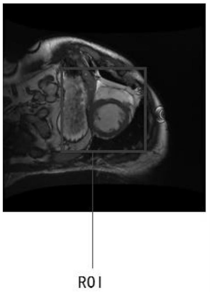
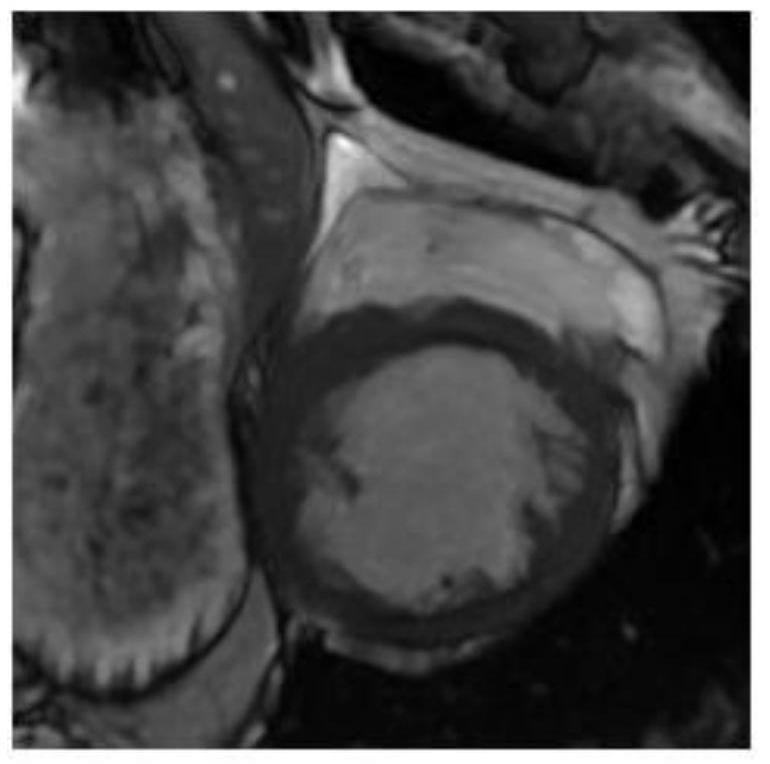
[0038] MRI images corresponding to each cardiac phase in the middle slice I MP Above, set the initial region of interest, calculate the maximum intensity projection image (MIP) of the initial region of interest on all cardiac phases (P cardiac phases), cluster and segment the maximum intensity projection image (MIP), and select the roundness The largest area A; the preferred clustering and segmentation method is the fuzzy C-means clustering method;

[0039]...


PUM


Abstract

The invention discloses a method for segmenting blood pools in end-diastolic images of cardiac functional magnetic resonance images. NP ; Magnetic resonance image I corresponding to each cardiac phase in the middle slice MP Above, calculate the maximum intensity projection image of the initial region of interest on all cardiac phases, cluster and segment the maximum intensity projection image, and select the area A with the largest roundness; calculate the magnetic resonance corresponding to each cardiac phase of the middle slice Image I MP The average gray value of the image in region A, the MRI image I with the largest average gray value MP is the MRI image I corresponding to the middle slice at the end of diastole MP(ED) ; Segment the MRI images from the middle slice to the apex and the bottom of the heart respectively to complete the end-diastolic MRI image I NP(ED) Segmentation of the blood pool in .

Description

【Technical field】 [0001] The invention relates to the field of medical images, in particular to the segmentation of cardiac functional magnetic resonance images. 【Background technique】 [0002] In recent years, the morbidity and mortality of cardiovascular diseases are increasing year by year. Nearly 6 million people in the United States are diagnosed with cardiovascular disease every year, while nearly 3 million people die of cardiovascular disease in my country. Cardiac function is an important indicator for the diagnosis of cardiovascular diseases, which can be achieved by analyzing the global and local characteristic parameters related to the left ventricle in the four-dimensional functional magnetic resonance image, such as: ventricular volume, ejection fraction, and myocardial wall thickness. The accuracy of the extraction of cardiac function feature parameters depends on the accurate localization of the left ventricular endocardium, and the computer-aided diagnosis s...

Claims


Application Information

Patent Timeline
Patent Type & Authority Patents(China)
IPC IPC(8): G06T7/11G06K9/62
CPCG06T2207/30048G06T2207/20104G06F18/23211
Inventor 姜娈凌姗李强
Owner SHANGHAI UNITED IMAGING HEALTHCARE
Why Eureka
Try Eureka