Porous bioceramic support and preparation method thereof

A technology for bioceramics and ceramic bodies, applied in the field of medical materials, can solve the problems of limiting the size and number of communicating pores, low communicating pores, and ineffective contact of porogens

Active Publication Date: 2020-11-27
SHENZHEN LANDO BIOMATERIALS
View PDF8 Cites 0 Cited by
  • Summary
  • Abstract
  • Description
  • Claims
  • Application Information

AI Technical Summary

Problems solved by technology

Liu Weijian et al. (Preparation and Characterization of Gradient Scaffold Materials for Tissue Engineering, Beijing University of Chemical Technology, 2006) used a stacking template of sodium chloride particles with different particle sizes. Although the template has a certain gradient structure, the sodium chloride particle It is square, and the uniformity of the interconnected pores formed after stacking is low. In addition, the polylactic acid and the porogen are mixed first and then injected into the mold. The pore structure of the obtained porous bioceramic scaffold is quite different from that of natural bone, which affects the application

Method used


Image

  • Porous bioceramic support and preparation method thereof
  • Porous bioceramic support and preparation method thereof
  • Porous bioceramic support and preparation method thereof

Examples


preparation example Construction

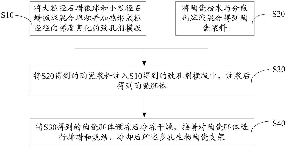
[0031] Such as figure 1 The preparation method of the porous bioceramic support of one embodiment shown, comprises the following steps:

[0032] S10, mixing and stacking the large-diameter paraffin microspheres and the small-diameter paraffin microspheres, and heating to form a porogen template with radial gradient changes in particle diameter.

[0033] The large-diameter paraffin microspheres can be spherical paraffin microspheres or ellipsoidal paraffin microspheres, and the small-diameter paraffin microspheres can be spherical paraffin microspheres or ellipsoidal paraffin microspheres.

[0034] The particle diameter of the paraffin wax microspheres with large particle diameter may be 600 μm-1200 μm, and the particle diameter of paraffin wax microspheres with small particle diameter may be 50 μm-600 μm.

[0035] In the porogen template, the mass ratio of large-diameter paraffin microspheres to small-diameter paraffin microspheres is 1:0.1-10.

[0036] The operation of mixi...

Embodiment 1

[0054] 1. Screen out paraffin microspheres with a particle size ranging from 600 μm to 1200 μm as large paraffin microspheres, and paraffin microspheres with a particle size ranging from 50 μm to 600 μm as small paraffin microspheres.

[0055] 2. Large-diameter paraffin microspheres are accumulated inside the cylindrical mold, and heated at 45°C for 15 minutes after flattening, so that the large-diameter paraffin microspheres are bonded to each other, and large-diameter paraffin microspheres are formed after cooling Template: small-diameter paraffin microspheres are deposited on the outer periphery of the cylindrical mold, flattened and withdrawn from the cylindrical mold, then heated at 45°C for 15 minutes, and cooled to form a porogen template with radial gradients in particle size. Wherein, the mass ratio of large-diameter paraffin microspheres and small-diameter paraffin microspheres is 2:1.

[0056] 3. Prepare an ammonium polyacrylate dispersant solution with a mass perce...

Embodiment 2

[0063] 1. Screen out paraffin microspheres with a particle size ranging from 600 μm to 1200 μm as large paraffin microspheres, and paraffin microspheres with a particle size ranging from 50 μm to 600 μm as small paraffin microspheres.

[0064] 2. Large-diameter paraffin microspheres are deposited inside the cylindrical mold, and then small-diameter paraffin microspheres are deposited on the outer periphery of the cylindrical mold. After flattening, the cylindrical mold is withdrawn, and then heated at 40°C for 30 minutes. After cooling A porogen template with radial gradient changes in particle size is formed. Wherein, the mass ratio of large-diameter paraffin microspheres and small-diameter paraffin microspheres is 1:1.

[0065] 3. Prepare a sodium polyacrylate dispersant solution with a mass percentage concentration of 1%, mix hydroxyapatite and β-tricalcium phosphate in a mass ratio of 1:1 to form a ceramic powder, and then press the ceramic powder: ammonium polyacrylate di...


PUM

PropertyMeasurementUnit
particle diameteraaaaaaaaaa

Abstract

The invention discloses a porous biological ceramic scaffold and a preparation method thereof. The preparation method comprises: mixing and stacking large particle size paraffin microspheres and small particle size paraffin microspheres, and heating to form a pore forming agent template with a characteristic of radial particle size gradient change; mixing ceramic powder and a dispersing agent solution to obtain a ceramic slurry; injecting the ceramic slurry into the pore forming agent template, and grouting to obtain a ceramic billet; and pre-freezing the ceramic billet, carrying out freeze drying, dewaxing and sintering the ceramic billet, and cooling to obtain the porous biological ceramic scaffold. According to the present invention, the large particle size paraffin microspheres and the small particle size paraffin microspheres are subjected to the mixing stacking, and the heating is performed to form the pore forming agent template with the characteristic of radial particle size gradient change, such that the pore structure of the prepared porous biological ceramic scaffold is close to the structure of the natural bone, and the prepared porous biological ceramic scaffold is the excellent bone repair material.

Description

technical field [0001] The invention relates to the field of medical materials, in particular to a porous bioceramic support and a preparation method thereof. Background technique [0002] Compared with the traditional bone repair solutions - autologous bone, allogeneic bone and xenogeneic bone materials, artificial bone repair materials have the advantages of no immune rejection, no risk of animal origin, low cost, wide range of sources, and exemption from secondary surgery for patients. It is considered to be one of the most promising bone repair materials. [0003] In order to make it easier for cells and blood vessels to grow into the material to promote the rapid formation of new bone tissue, artificial bone repair materials are often prepared in the shape of a connected porous scaffold that mimics the natural bone structure. The natural bone structure is a radial gradient structure with a loose interior and a relatively dense exterior. Holes of different sizes are sui...

Claims


Application Information

Patent Timeline
Patent Type & Authority Patents(China)
IPC IPC(8): C04B38/06A61L27/56A61L27/10
CPCA61L27/10A61L27/56C04B38/061C04B38/0054
Inventor 潘洋郭远军谭荣伟佘振定
Owner SHENZHEN LANDO BIOMATERIALS
Why Eureka
Try Eureka