A method for producing bone and soft tissue X-ray imaging images

A production method and soft tissue technology, which is applied in the field of medical surgery and medical teaching images, can solve problems that plague technicians, are difficult to achieve, and cannot produce images showing the spatial positions of bones and soft tissues, so as to improve teaching quality and achieve significant social benefits Effect

Active Publication Date: 2018-12-18
THE THIRD HOSPITAL OF HEBEI MEDICAL UNIV
View PDF3 Cites 0 Cited by
  • Summary
  • Abstract
  • Description
  • Claims
  • Application Information

AI Technical Summary

Problems solved by technology

But the fundamental problem that exists at present is that X-rays cannot develop soft tissues such as muscles, tendons, ligaments, blood vessels, and nerves during fluoroscopy, but can only develop bones, so it is impossible to display bones and bones in various parts of the human body through X-ray fluoroscopy. The image of the spatial position of the soft tissue makes it difficult for people to realize this idea
How to display human soft tissues through X-rays, and then make images showing the spatial positions of bones and soft tissues in various parts of the human body has become a problem that has plagued technicians for a long time, and has not been resolved so far

Method used


Image

  • A method for producing bone and soft tissue X-ray imaging images
  • A method for producing bone and soft tissue X-ray imaging images

Examples


Embodiment Construction

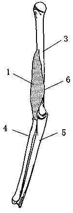
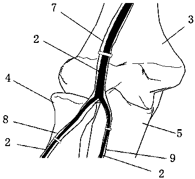
[0023] The present invention is a method for making X-ray imaging images that can display the mutual positional relationship between bones and soft tissues. relationship, and then produce a three-dimensional stereoscopic image, which provides a direct reference standard for minimally invasive surgery X-ray operations, and can be used in medical teaching to improve the quality of teaching.

[0024] The core of this method is the technique of developing soft tissue under X-ray, which should meet the following requirements:

[0025] First of all, muscles, tendons, ligaments, joint capsules and neurovascular tissues can be visualized under X-ray, and the X-ray films taken can clearly show the distribution and running characteristics of soft tissue structures, and can clearly show the relationship between soft tissues and bones;

[0026] Secondly, the anatomical position is accurate, which has guiding significance for clinical practice;

[0027] Again, it is easy to understand, ea...


PUM


Abstract

The invention relates to a method for producing X-ray imaging images of bones and soft tissues, which belongs to the technical field of medical operations and medical teaching images, and is used for producing X-ray imaging images capable of displaying the mutual positional relationship between bones and soft tissues. The technical solution is: the present invention uses metal foil or wire mesh that can be developed under X-rays and can pass through X-rays to cover the muscles, tendons, ligaments, and joint capsules that need to be developed, and at the same time stick medical barium wires to blood vessels and nerves. In general, X-ray imaging is performed on the developed area according to the common projection angles in minimally invasive surgery, and the imaging data of the spatial position between bones and muscles, tendons, ligaments, joint capsules, blood vessels, and nerves can be obtained. The invention is the first of its kind in medical treatment and medical teaching, which solves the long-term unsolved problem of displaying human soft tissues through X-rays, provides direct reference standards for X-ray operations of minimally invasive surgery, and provides scientific and technological support for the development of minimally invasive surgery in orthopedics. And it can be used in medical teaching to improve the teaching quality.

Description

technical field [0001] The invention relates to a method for making an X-ray developing image capable of displaying the mutual positional relationship between bones and soft tissues, and belongs to the technical fields of medical operations and medical teaching images. Background technique [0002] Minimally invasive surgery for fracture treatment is an unstoppable trend in the development of orthopedics. Familiarity with the anatomical structures around the bone, such as the distribution of muscles, tendons, ligaments, joint capsules, and neurovascular, is an important condition to ensure the safe completion of minimally invasive surgery. With the continuous expansion of the scope of orthopedic minimally invasive surgery, it is increasingly important to have a clear understanding of the anatomical structure between bones and soft tissues in various parts of the human body, so as to prevent doctors from not fully understanding important anatomical structures during the opera...

Claims


Application Information

Patent Timeline
Patent Type & Authority Patents(China)
IPC IPC(8): G03B42/02G09B5/02
CPCG03B42/026G09B5/02
Inventor 张英泽陈霄付蕾李彦森陈伟邢恩增刘涛
Owner THE THIRD HOSPITAL OF HEBEI MEDICAL UNIV
Why Eureka
Try Eureka