
Assessment of Myocardial Infarction by Real-Time Ultrasound Strain Imaging

A technology of imaging and ultrasonic diagnosis, applied in the directions of ultrasonic/sonic/infrasonic image/data processing, ultrasonic/sonic/infrasonic diagnosis, ultrasonic/sonic/infrasonic Permian technology, etc., which can solve the lack of sensitivity in diagnosing local cardiac function, etc. question

Active Publication Date: 2020-06-30
KONINKLJIJKE PHILIPS NV
View PDF7 Cites 0 Cited by
  • Summary
  • Abstract
  • Description
  • Claims
  • Application Information

AI Technical Summary

Problems solved by technology

While useful, they cannot be used to localize infarcts and reliably visualize or delineate ischemic regions
These applications lack the sensitivity required to diagnose regional cardiac function
Furthermore, only one strain image can be produced for a cardiac cycle since strain is determined by tracking myocardial motion throughout the cardiac cycle, or at least its systolic phase

Method used


Image

  • Assessment of Myocardial Infarction by Real-Time Ultrasound Strain Imaging
  • Assessment of Myocardial Infarction by Real-Time Ultrasound Strain Imaging
  • Assessment of Myocardial Infarction by Real-Time Ultrasound Strain Imaging

Examples


Embodiment Construction

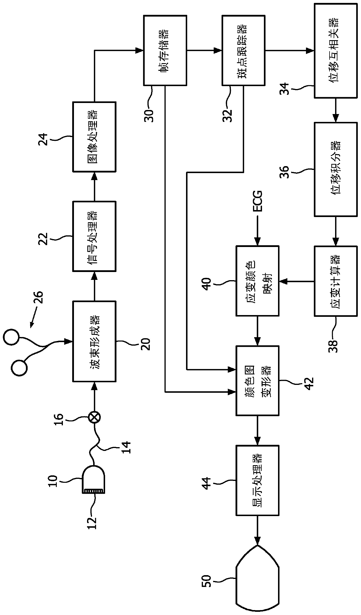
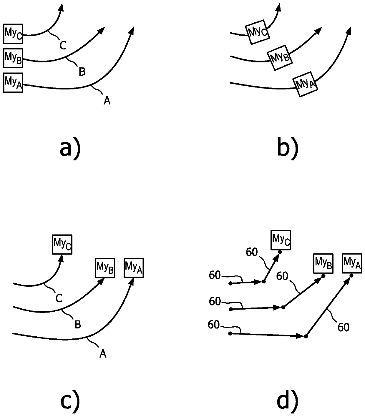
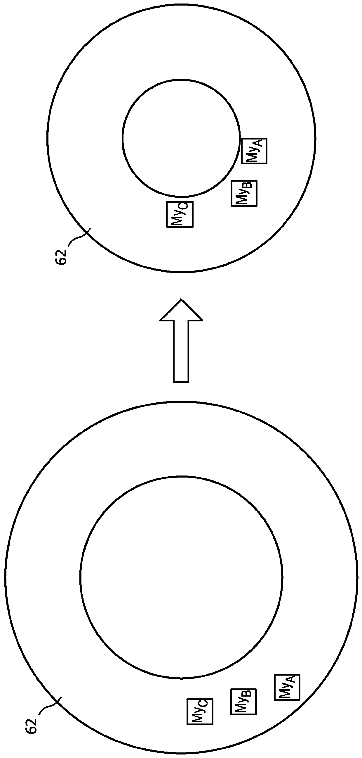
[0015] In accordance with the principles of the present invention, an ultrasonic diagnostic imaging system is described that is capable of imaging the heart at high frame rates and calculating strain in localized regions of the myocardium. For each pixel on the image, a strain parameter representing the local strain is determined, and then these pixel values ​​are spatially mapped to the anatomical image. The strain map is then fitted to the first image of the next cardiac cycle and displayed as a parametric color overlay on the image frame of the next cycle of the cardiac image. The color overlay is deformed to fit continuously across each heart image as the image changes as the myocardium contracts and relaxes. Thus, the heart, its spatial strain changes, and corresponding contractile properties are displayed to the user in real time.

[0016] In some aspects, the present invention provides an ultrasonic diagnostic imaging system for real-time strain imaging. The ultrasoun...


PUM


Abstract

Real-time strain imaging is provided by acquiring a sequence of cardiac image frames and estimating displacement in the myocardium over the cardiac cycle. Displacement can be estimated using speckle tracking and used to calculate the strain on the myocardium. The colormap includes strain values. During the next cardiac cycle, warping the color map to fit the myocardium in each image frame, and displaying the warped color map as a new cardiac cycle while displaying the warped color map in real time The myocardium is overlaid in color for each image. A new colormap is also generated on the new heart cycle for use with subsequent heart cycles. An ultrasound system performing real-time strain imaging is also described.

Description

[0001] priority claim [0002] This application claims priority to US Provisional Application No. 62 / 109209, filed January 29, 2015, which is incorporated herein by reference. technical field [0003] The present invention relates to ultrasonic diagnostic imaging systems, and more particularly to the use of ultrasonic strain imaging to assess ischemic regions of a heart that has suffered an infarction. Background technique [0004] One use of cardiac ultrasound is to evaluate the heart of a patient suffering from an infarction. For example, it would be desirable to be able to image coronary arteries to look for blockages that may cause ischemic events. However, ultrasound typically cannot directly visualize the coronary arteries due to their motion, location outside the heart, proximity to the lungs, and other characteristics. Therefore, arterial function is indirectly diagnosed by evaluating myocardial wall motion using ultrasound. Abnormal or asynchronous wall motion in...

Claims


Application Information

Patent Timeline
Patent Type & Authority Patents(China)
IPC IPC(8): A61B8/08A61B8/00
CPCA61B8/0883A61B8/485A61B8/5246A61B8/461A61B8/543
Inventor A·V·帕蒂尔A·英格尔K·E·蒂勒
Owner KONINKLJIJKE PHILIPS NV
Learn More