
Detection method and device of image

A detection method and image technology, applied in the medical field, can solve problems such as low precision, large error, and time-consuming

Active Publication Date: 2017-11-21
UNIV OF ELECTRONICS SCI & TECH OF CHINA
View PDF3 Cites 13 Cited by
  • Summary
  • Abstract
  • Description
  • Claims
  • Application Information

AI Technical Summary

Problems solved by technology

This is a very difficult task. Once the hand holding the measuring stick shakes slightly, it will affect the image quality of the tumor
Therefore, the existing manual measurement and marking methods need to consume a lot of time, the labor cost is high, the accuracy is relatively low, and the error is relatively large

Method used


Image

  • Detection method and device of image
  • Detection method and device of image
  • Detection method and device of image

Examples


no. 1 example

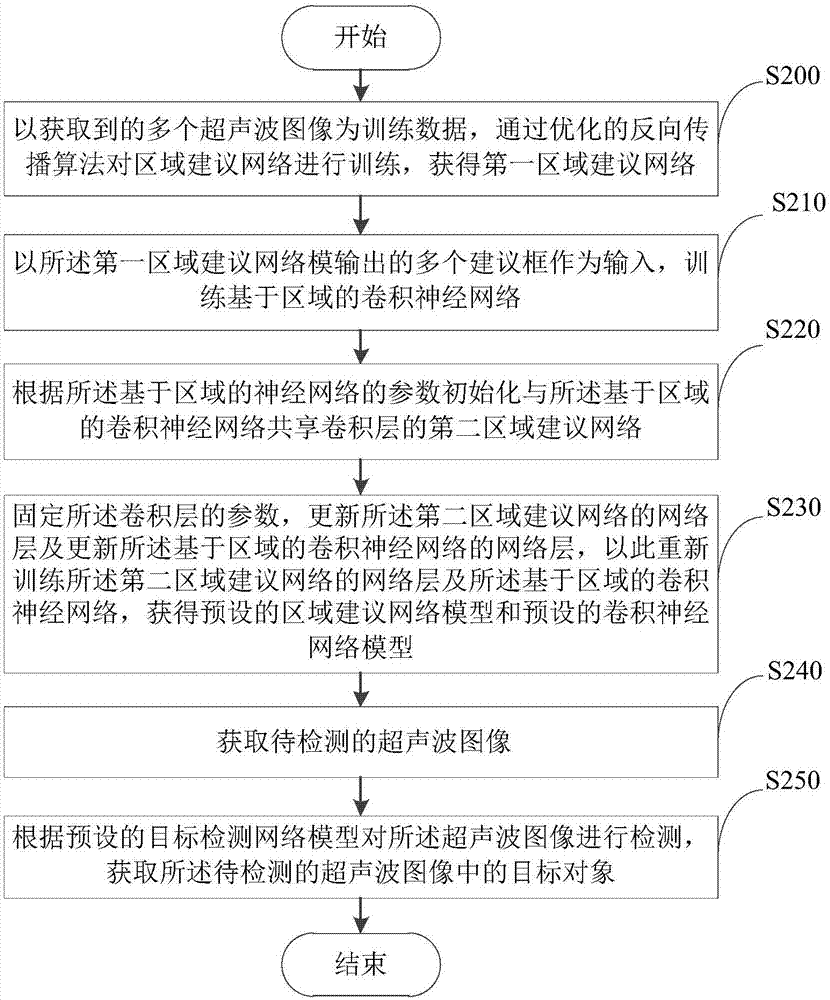
[0030] see figure 2 , the embodiment of the present invention provides an image detection method, the method includes step S200, step S210, step S220, step S230, step S240 and step S250.

[0031] Step S200: Using the multiple acquired ultrasound images as training data, train the region proposal network through an optimized backpropagation algorithm to obtain a first region proposal network.

[0032] In this embodiment, the plurality of ultrasonic images may all be breast ultrasonic images, which may come from the ImageNet model.

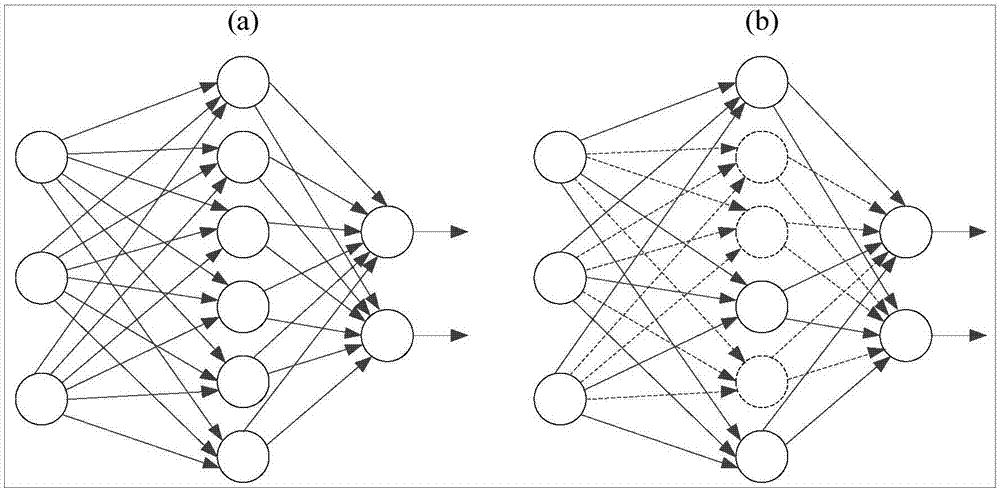
[0033] Based on step S200, further, using multiple acquired ultrasonic images as training data, the region proposal network is modified by using Dropout, and the modified region proposal network is trained by an optimized backpropagation algorithm to obtain the first Regional networking.

[0034] The cost function of the optimized backpropagation algorithm is based on adding an L1 regular term or an L2 regular term to the original cost function. ...

no. 2 example

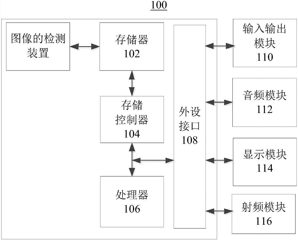
[0078] see Figure 6 , the embodiment of the present invention provides an image detection device 300 , the device 300 shown includes a suggestion network training unit 310 , a convolutional neural network training unit 320 , an initialization unit 330 , an update unit 340 , an acquisition unit 350 and a detection unit 360 .

[0079] The suggested network training unit 310 is configured to use the acquired multiple ultrasound images as training data to train the region proposed network through an optimized backpropagation algorithm to obtain a first region proposed network.

[0080] The suggested network training unit 310 includes a suggested network training subunit 311 .

[0081] The proposed network training subunit 311 is configured to use the acquired multiple ultrasonic images as training data, use Dropout to modify the region proposed network, and train the modified region proposed network through an optimized backpropagation algorithm to obtain The first area establis...


PUM


Abstract

The embodiment of the invention provides a detection method and device of an image, and relates to the field of medical technology. The method includes: acquiring the to-be-detected ultrasound image; and then detecting the to-be-detected ultrasound image according to a preset target detection network model to acquire a target object in the to-be-detected ultrasound image. Therefore, automatically detecting the target object in the ultrasound image is realized, operations are simple, the precision is high, and consumed time is short.

Description

technical field [0001] The present invention relates to the field of medical technology, in particular to an image detection method and device. Background technique [0002] At present, the detection and measurement of tumors in the ultrasound images to be detected in the hospital are carried out with the experience of doctors. But the marking and measurement of tumors is a very troublesome thing, because it requires the skill of doctors. When the doctor thinks that a better tumor angle can be presented on the B-ultrasound image, one hand needs to hold the measuring stick for a long time and keep it fixed, and the other hand operates on the machine for measurement. This is a very difficult task. Once the hand holding the measuring stick shakes slightly, it will affect the image quality of the tumor. Therefore, the existing manual measurement and marking methods need to consume a lot of time, have high labor costs, relatively low precision, and relatively large errors. Co...

Claims


Application Information

Patent Timeline
IPC IPC(8): G06T7/00G06N3/04G06N3/08
CPCG06T7/0012G06T2207/10132G06T2207/20081G06T2207/20084G06T2207/30068G06T2207/30096
Inventor 杨国武秦晓明曹占涛曾安军聂婧梁涛董俊文吕凤毛林晨张艳张菊玲
Owner UNIV OF ELECTRONICS SCI & TECH OF CHINA
Learn More