CT lung nodule detection system based on 3D-Unet

A detection system and technology for pulmonary nodules, applied in image enhancement, image analysis, image data processing, etc., can solve the problems of doctors' visual fatigue, increased workload, missed diagnosis, etc., achieve high detection accuracy, solve heavy workload, The effect of improving the recognition rate

Active Publication Date: 2018-10-12
四川元匠科技有限公司
View PDF12 Cites 30 Cited by
  • Summary
  • Abstract
  • Description
  • Claims
  • Application Information

AI Technical Summary

Problems solved by technology

[0003] The traditional lung nodule detection method is through CT scanning, and the doctor manually marks the lung nodules. The principle of CT is tomographic scanning. The complete lung CT image generated by one examination of a case often contains hundreds of pieces, which greatly increases the workload and is easy to detect. It causes doctors’ visual fatigue and increases the probability of misdiagnosis and missed diagnosis. At the same time, due to the complex structure of the lungs, the characteristics of pulmonary nodules on CT images are similar to those of pulmonary blood vessels and bronchial sections, which cannot be effectively distinguished based on CT values. Due to the relatively random distribution of pulmonary nodules, adhesions to the lung cavity membrane and blood vessels often occur, and the morphological differences between nodules are relatively large. Even experienced radiologists can hardly make a quick decision. diagnosis

Method used


Image

  • CT lung nodule detection system based on 3D-Unet
  • CT lung nodule detection system based on 3D-Unet
  • CT lung nodule detection system based on 3D-Unet

Examples


Embodiment Construction

[0036] Further describe the technical scheme of the present invention in detail below in conjunction with accompanying drawing:

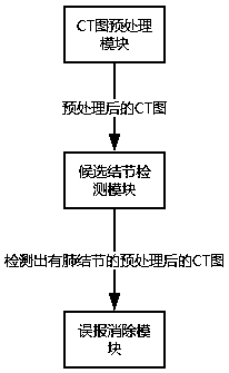
[0037] Such as figure 1 As shown, a 3D-Unet-based CT image pulmonary nodule detection system includes sequentially connected CT image input and preprocessing modules, candidate nodule detection modules, and false alarm elimination modules.

[0038] Among them, when the CT image is taken, the edge of the machine or the patient's bone will also be photographed, which is noise for the neural network and is not conducive to processing, so the preprocessing is to make the lung as much as possible. Keep it, and remove some irrelevant things at the same time.

[0039] Specifically, the CT image input and preprocessing module is used to read the chest CT image in DICOM format and save the image information into a numpy array, obtain the distance and origin information of the CT image, and perform lung volume calculation on the CT image. segmentation.

[...


PUM


Abstract

The invention discloses a CT lung nodule detection system based on 3D-Unet. The system includes a CT image input and preprocessing module, a candidate nodule detection module and a false-reporting elimination module which are sequentially connected. The CT image input and preprocessing module is used for reading a chest CT graph, obtaining spacing and origin information of the CT graph, and carrying out lung volume segmentation on the CT graph. The candidate nodule detection module is used for inputting the image, of which preprocesing is completed, into a Unet network, and obtaining a location of a candidate lung nodule. The Unet network includes sixteen residual blocks, two path units, four maximum-pooling units, one preparation unit, one probabilistic neuron failure unit and one outputunit. According to the system, automatic detection of the lung nodule is realized, problems of high doctor workloads and high misdiagnosis rates in lung nodule diagnosis are solved, an effect of detecting the lung nodule by the 3D-Unet is realized, more contextual semantic information in the CT image is utilized, and detection accuracy is higher.

Description

technical field [0001] The invention relates to a 3D-Unet-based CT image pulmonary nodule detection system. Background technique [0002] Lung cancer is one of the malignant tumors with the fastest growing morbidity and mortality, and the greatest threat to the health and life of the population, with a mortality rate as high as 91.6%. Pulmonary nodules are the most common form of early lung cancer. The detection of lung cancer ultimately comes down to the detection of pulmonary nodules, and CT imaging can directly display and observe the lesions. [0003] The traditional lung nodule detection method is through CT scanning, and the doctor manually marks the lung nodules. The principle of CT is tomographic scanning. The complete lung CT image generated by one examination of a case often contains hundreds of pieces, which greatly increases the workload and is easy to detect. It causes doctors’ visual fatigue and increases the probability of misdiagnosis and missed diagnosis. A...

Claims


Application Information

Patent Timeline
Patent Type & Authority Applications(China)
IPC IPC(8): G06T7/00
CPCG06T7/0012G06T2207/10081G06T2207/20081G06T2207/20084G06T2207/30064
Inventor 臧宇航朱安婕郑德生张雪吉普照
Owner 四川元匠科技有限公司
Why Eureka
Try Eureka