A Method for Calculating Cardiothoracic Ratio in Medical Images

A medical image and cardiothoracic ratio technology, applied in the field of medical image processing, can solve the problems that cannot meet the requirements of high precision, real-time and automation at the same time

Active Publication Date: 2020-11-20
SHANGHAI UNITED IMAGING HEALTHCARE
View PDF5 Cites 0 Cited by
  • Summary
  • Abstract
  • Description
  • Claims
  • Application Information

AI Technical Summary

Problems solved by technology

The above methods for calculating cardiothoracic ratio cannot meet the requirements of high precision, real-time and automation at the same time

Method used


Image

  • A Method for Calculating Cardiothoracic Ratio in Medical Images
  • A Method for Calculating Cardiothoracic Ratio in Medical Images
  • A Method for Calculating Cardiothoracic Ratio in Medical Images

Examples


Embodiment Construction

[0041] The present invention will be further described below in conjunction with the accompanying drawings and embodiments.

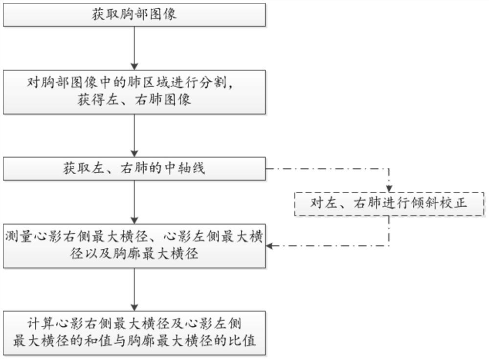
[0042] see figure 1 , a method for calculating cardiothoracic ratio in a medical image according to an embodiment of the present invention, comprising the following steps:

[0043] A chest image is obtained, the medical image is obtained by an X-ray device, a CT device or an MR device.

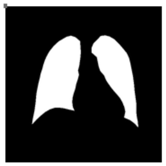
[0044] Segment the lung area in the chest image to obtain left and right lung images;

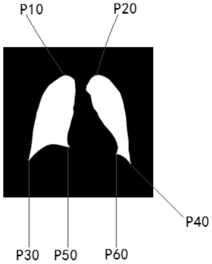
[0045] Obtain the central axis of the left and right lungs;

[0046] Automatically measure (calculate) the largest transverse diameter on the right side of the heart shadow, the largest transverse diameter on the left side of the heart shadow, and the largest transverse diameter of the thorax;

[0047] Calculate the ratio of the sum of the maximum transverse diameter on the right side of the heart shadow and the maximum transverse diameter on the left side of the heart shadow to t...


PUM


Abstract

The invention discloses a method for cardio-thoracic proportion calculation of a medical image. The method includes steps of acquiring a thorax image; performing segmentation on a lung area in the thorax image and acquiring a left lung image and a right lung image; acquiring the central axis of the left lung and the right lung; measuring a podoid right side maximal cross diameter, a podoid left side maximal cross diameter and a thorax maximal cross diameter; calculating the proportion between the sum of the podoid right side maximal cross diameter and the podoid left side maximal cross diameter and the thorax maximal cross diameter. According to the invention, on the basis of lung segmentation, the podoid maximal cross diameter and the thorax maximal cross diameter are measured automatically and the cardio-thoracic proportion is calculated.

Description

【Technical field】 [0001] The invention relates to the field of medical image processing, in particular to a calculation method of a cardiothoracic ratio (CTR) in a chest image. 【Background technique】 [0002] Medical imaging is an auxiliary diagnostic technology that helps doctors understand the health status of patients. With the development of science and technology, various medical imaging equipment, such as X-ray equipment, DR equipment, CT equipment, and MR equipment, have been created and widely used, thereby helping Doctors understand the internal tissue structure of the examined body, which greatly improves the diagnostic level of modern medicine. Digital X-ray film (DR image) is relatively simple to operate, easy to use and low in cost, and has been widely used. [0003] In clinical practice, chest imaging is often used to evaluate the heart and lung regions for conditions such as cardiac hypertrophy. There are many diagnostic indicators for assessing cardiac hype...

Claims


Application Information

Patent Timeline
Patent Type & Authority Patents(China)
IPC IPC(8): G06T7/62G06T7/66G06T7/11G06T7/13G06T7/136
CPCG06T2207/10081G06T2207/10088G06T2207/10116G06T2207/30048
Inventor 丛琳霍士虎陈刚李强
Owner SHANGHAI UNITED IMAGING HEALTHCARE
Why Eureka
Try Eureka