Breast region segmentation and calcification detection method in mammography images

A region segmentation and detection method technology, applied in the field of biomedical imaging and biomedical detection, can solve the problems of affecting the analysis results, different breast shapes, small projected area of ​​calcification points, etc., to achieve obvious economic and social benefits, broad economic and Social benefits and the effect of improving image processing efficiency

Active Publication Date: 2021-04-27
FUJIAN NORMAL UNIV
View PDF8 Cites 0 Cited by
  • Summary
  • Abstract
  • Description
  • Claims
  • Application Information

AI Technical Summary

Problems solved by technology

[0004] For the application of mammography image processing and analysis, manual recognition can only qualitatively estimate the boundary between the breast and the background and the clearer boundary between the breast and chest muscles, which has been difficult to meet the accuracy and speed requirements of breast morphology analysis. Traditional mammography image processing and analysis methods also have shortcomings that seriously affect the analysis results: the breast itself has various shapes, and it is difficult to use traditional methods based on morphological models to segment various tissues; the nucleus, cytoplasm and extracellular matrix The distribution of calcification is uneven, it is difficult to use traditional texture features to generalize and analyze the image, and the calculation of local texture features is large, and the efficiency is not high; the projected area of ​​calcification is small, and the contrast with the surrounding tissue is not high
The above shortcomings lead to inaccurate division of boundaries between breast and image background and chest muscles, as well as inaccurate detection of breast calcification points, resulting in errors in the statistical analysis of different types of tissues in mammography images, which seriously affect the discrimination of mammography image processing Accuracy and processing speed

Method used


Image

  • Breast region segmentation and calcification detection method in mammography images
  • Breast region segmentation and calcification detection method in mammography images
  • Breast region segmentation and calcification detection method in mammography images

Examples


Embodiment Construction

[0085] In order to make the features and advantages of this patent more obvious and easy to understand, the following special examples are described in detail as follows:

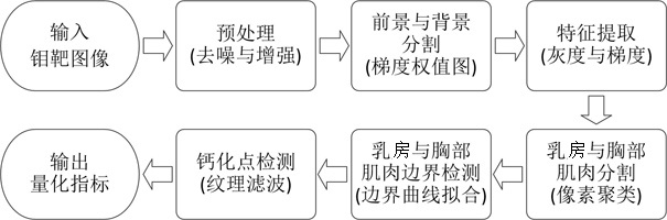
[0086] Such as figure 1 Shown, the embodiment of the present invention comprises the following steps:

[0087] Step 1: Preprocessing the original image of mammography, including image denoising and enhancement, to obtain a grayscale image with enhanced pixel signal and clearer boundaries of various tissues;

[0088] Step 2: Calculating the corresponding gray gradient weight image for the preprocessed mammography image;

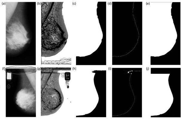
[0089] Step 3: Perform erosion and expansion operations on the closed area of ​​the gray-scale gradient weight image, check the inflection point of the boundary between the upper breast and the adhesion artificial interference, remove the artificial interference in the image, and obtain an image containing only the breast and chest muscles the border between the foreground area and the im...


PUM


Abstract

The present invention proposes a method for breast region segmentation and calcification point detection in mammography images. The method uses an image gradient weight calculation method based on neighboring pixels, so as to realize rapid removal of artificial interference in mammography images, and Due to the use of the image segmentation algorithm based on pixel clustering, the initial segmentation of breast and chest muscles is realized. In addition, due to the use of the straight line detection algorithm based on Hough transform and the curve fitting algorithm based on polynomial, it can accurately detect and fit the breast boundary. , so that the method of the present invention is adopted to significantly improve the marking accuracy of the boundary between the breast, the background and the chest muscles in the mammography image, and finally, due to the use of texture filtering to detect calcification points in the breast, the breast area calibration and The accuracy of calcification point detection can realize automatic segmentation and calibration of breast regions and automatic detection and marking measurement of calcification points.

Description

technical field [0001] The invention relates to the fields of biomedical imaging and biomedical detection, in particular to a method for segmenting breast regions and detecting calcification points in mammography images based on pixel clustering and texture filtering. Background technique [0002] The full name of mammography mammography mammography, also known as molybdenum palladium examination, is the first choice, the simplest and most reliable non-invasive detection method for the diagnosis of breast diseases. It is relatively painless, easy to implement, and has high resolution. , good reproducibility, the retained images can be compared before and after, and are not limited by age and body shape. It has been used as a routine inspection method at present. As a relatively non-invasive examination method, mammography can comprehensively and correctly reflect the general anatomical structure of the whole breast, observe the influence of various physiological factors such...

Claims


Application Information

Patent Timeline
Patent Type & Authority Patents(China)
IPC IPC(8): G06T7/00G06T7/11G06T7/13G06T7/194G06T7/187
CPCG06T7/0012G06T2207/10081G06T2207/20032G06T2207/20081G06T2207/30068G06T7/11G06T7/13G06T7/187G06T7/194
Inventor 时鹏钟婧
Owner FUJIAN NORMAL UNIV
Why Eureka
Try Eureka