Medical image display method and apparatus

A medical image and display method technology, applied in the field of medical images, can solve the problems of time-consuming, complex multi-window display settings, etc., and achieve the effect of saving operation time

Inactive Publication Date: 2018-04-06
SHANGHAI UNITED IMAGING HEALTHCARE
View PDF3 Cites 12 Cited by
  • Summary
  • Abstract
  • Description
  • Claims
  • Application Information

AI Technical Summary

Problems solved by technology

[0004] In view of this, the purpose of the present invention is to propose a medical image display method and device to solve the complex and time-consuming technical problems in the multi-window display of medical images in the prior art

Method used


Image

  • Medical image display method and apparatus
  • Medical image display method and apparatus
  • Medical image display method and apparatus

Examples


Embodiment 1

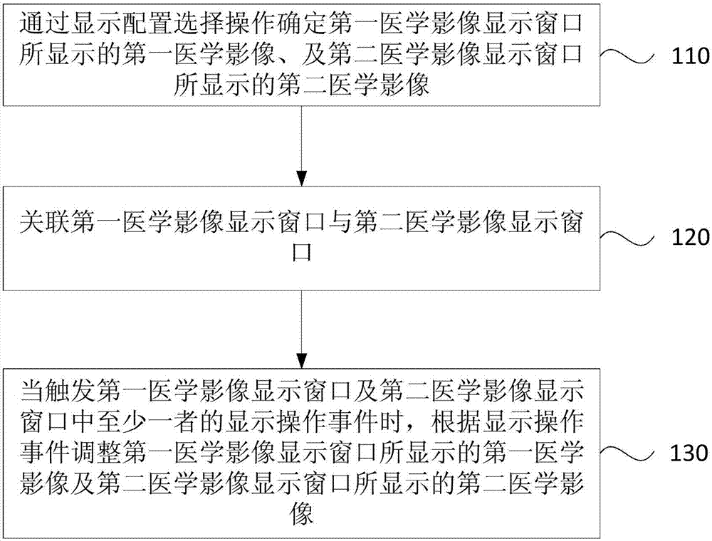
[0035] figure 1 It is a schematic flowchart of a medical image display method provided in Embodiment 1 of the present invention. The method is applicable to the application scene of medical image display operation, and the method can be executed by a medical image display device. The device can be realized by software and / or hardware. Such as figure 1 As shown, the method includes:

[0036] Step 110: Determine the first medical image displayed in the first medical image display window and the second medical image displayed in the second medical image display window through a display configuration selection operation.

[0037] Medical imaging is an image of the internal tissue of the human body or a part of the human body obtained in a non-invasive manner for medical treatment or medical research. Commonly used medical imaging techniques include: angiography (or angiography), cardiovascular angiography, CT, mammography, positron emission tomography, magnetic resonance imagi...

Embodiment 2

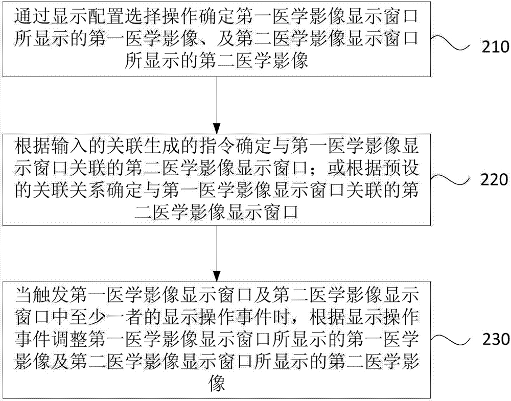
[0049] figure 2 It is a schematic flowchart of a medical image display method provided by Embodiment 2 of the present invention. Optimization is performed on the basis of the above embodiments, and the associating of the first medical image display window and the second medical image display window is specifically described. Such as figure 2 As shown, the method includes:

[0050] Step 210: Determine the first medical image displayed in the first medical image display window and the second medical image displayed in the second medical image display window through a display configuration selection operation.

[0051] Step 220: Determine the second medical image display window associated with the first medical image display window according to the input generated instruction; or determine the second medical image display window associated with the first medical image display window according to the preset association relationship .

[0052] To establish an association rela...

Embodiment 3

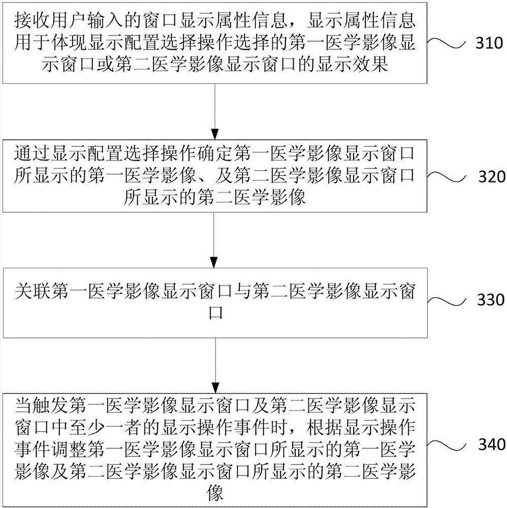
[0067] image 3 It is a schematic flowchart of a medical image display method provided by Embodiment 3 of the present invention. The optimization is performed on the basis of the above-mentioned embodiments, and the generation of the display configuration is described in detail. Such as image 3 As shown, the method includes:

[0068] Step 310: Receive window display attribute information input by the user, the display attribute information is used to reflect the display effect of the first medical image display window or the second medical image display window selected by the display configuration selection operation.

[0069] Exemplarily, the display attributes may include: displayed content and a displayed manner. For example: two-dimensional or three-dimensional; various attributes related to the displayed image, such as the resolution of the displayed content and the color configuration of the displayed content.

[0070] Exemplarily, the user can write the values ​​of...


PUM


Abstract

Embodiments of the invention disclose a medical image display method and apparatus. The method comprises the steps of confirming a first medical image displayed by a first medical image display windowand a second medical image displayed by a second medical image display window through display configuration selection operation; associating the first medical image display window with the second medical image display window; and when a display operation event of at least one of the first medical image display window and the second medical image display window is triggered, adjusting the first medical image displayed by the first medical image display window and the second medical image displayed by the second medical image display window according to the display operation event. The technical problems of complex setting and long time of medical image multi-window display in the prior art are solved.

Description

technical field [0001] The invention relates to the technical field of medical images, in particular to a medical image display method and device. Background technique [0002] When using various medical scanning equipment, such as ultrasound, computer tomography (Computed Tomography, CT), magnetic resonance imaging (Magnetic Resonance Imaging, MRI), X-ray to scan the patient, the doctor uses the configuration of the medical scanning equipment The display device observes the scanning images and makes judgments based on the images. At present, single-window or multi-window display scan images are usually used. For multi-window display, doctors can compare the different images displayed in each window to better judge the lesion. [0003] In the process of realizing the present invention, the inventor found the following technical problem: when using multi-windows to display medical scan images, since each window is displayed independently, the doctor needs to set the display...

Claims


Application Information

Patent Timeline
Patent Type & Authority Applications(China)
IPC IPC(8): G06F3/14G06F3/0484
CPCG06F3/04845G06F3/1423
Inventor 罗雪
Owner SHANGHAI UNITED IMAGING HEALTHCARE
Why Eureka
Try Eureka