
Construction and application of optical detection method of biological macromolecules related to intracellular colloid osmotic pressure and related drug screening method

A colloidal osmotic pressure, biomacromolecule technology, applied in biological testing, color/spectral property measurement, material analysis by optical means, etc., can solve problems such as less attention to intracellular mechanical activity

Active Publication Date: 2021-02-02
NANJING UNIVERSITY OF TRADITIONAL CHINESE MEDICINE
View PDF3 Cites 1 Cited by
  • Summary
  • Abstract
  • Description
  • Claims
  • Application Information

AI Technical Summary

Problems solved by technology

[0002] Chemical, electrical and mechanical activities are the basic forms of cell life activities. Although the former two have been studied in depth, intracellular mechanical activities have received little attention, which is a blind spot in the current cell research field

Method used


Image

  • Construction and application of optical detection method of biological macromolecules related to intracellular colloid osmotic pressure and related drug screening method
  • Construction and application of optical detection method of biological macromolecules related to intracellular colloid osmotic pressure and related drug screening method
  • Construction and application of optical detection method of biological macromolecules related to intracellular colloid osmotic pressure and related drug screening method

Examples


Embodiment 1

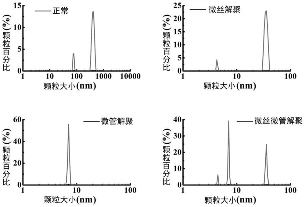
[0122] Determination of Myosin or Tubulin Particles and Their Particle Aggregates with Zetasizer Nano ZS90 Based on the Principle of Dynamic Light Scattering

[0123] It should be noted that the inventor intends to use this example to illustrate the operation method and principle of the nanometer particle size analyzer to observe myosin or tubulin particles and their particle aggregates, and is not limited to the Zetasizer Nano ZS90 instrument, nor It is limited to actin or tubulin and its particle aggregates, but is applicable to all nanometer particle size analyzers based on the principle of dynamic light scattering and all proteins and other macromolecules. The above examples are only used to illustrate the technology of the present invention Although the present invention has been described in detail with reference to preferred embodiments, those of ordinary skill in the art should understand that the technical solutions of the present invention can be modified or equivalen...

Embodiment 2

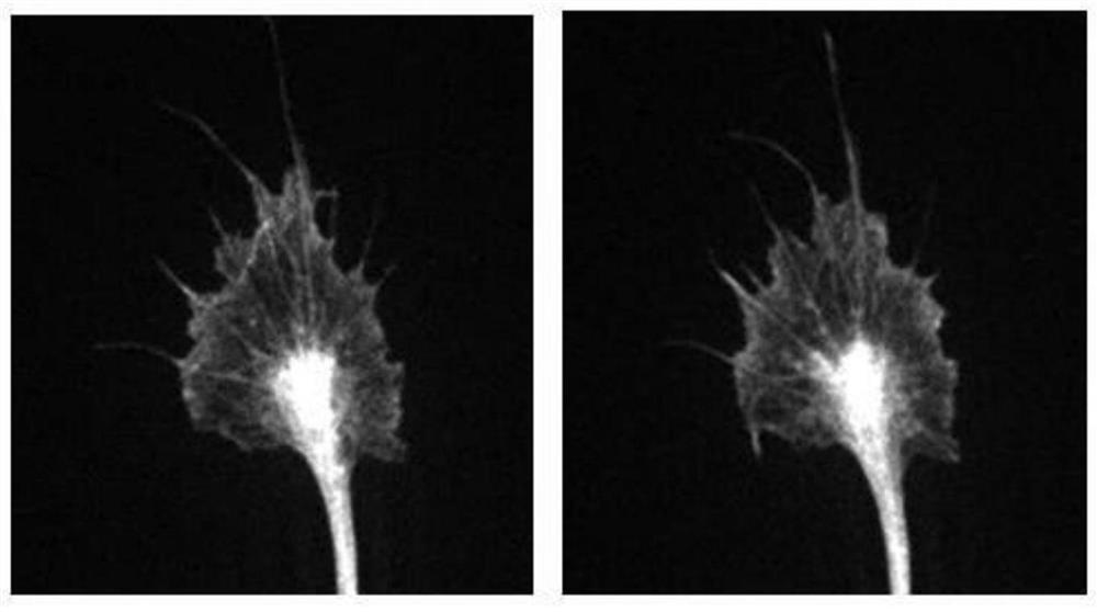
[0138] Quantity and distribution of myosin or tubulin granules and their granule aggregates in cells observed by total internal reflection dark-field microscopy

[0139] It should be noted that the present inventor intends to illustrate the operating method and principle of dark-field microscope observation of myosin or tubulin particles and their particle aggregates through this example, and is not limited to actin or tubulin and their aggregates. Particle aggregates, but applicable to all proteins and other macromolecules, the above embodiments are only used to illustrate the technical solutions of the present invention without limitation, although the present invention has been described in detail with reference to the preferred embodiments, those of ordinary skill in the art It should be understood that the technical solutions of the present invention can be modified or equivalently replaced without departing from the spirit and scope of the technical solutions of the prese...

Embodiment 3

[0151] The concentration of actin or tubulin granules is determined based on the specific absorbance of specific proteins.


PUM


Abstract

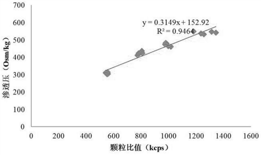
The invention relates to a biological macromolecule optical detection method related to intracellular colloid osmotic pressure, and construction and applications of a drug screening method related tothe biological macromolecule optical detection method. According to the present invention, the size, the shape, the position and the number of myosin, tubulin and other biological macromolecule particles or particle aggregates rich in cells or the density and the strength of protein markers are subjected to optical detection or imaging detection so as to evaluate the change of the intracellular colloid osmotic pressure and the regional mapping; with the optical detection method for the biological macromolecules related to intracellular colloid osmotic pressure, the change of the intracellularcolloid osmotic pressure can be analyzed, and the compositions and the functions of the crystal osmotic pressure and the colloid osmotic pressure in the intracellular colloid osmotic pressure can be analyzed by combining the method and the osmometer.

Description

technical field [0001] The invention relates to a biomacromolecule optical detection method, in particular to the construction and application of a biomacromolecule optical detection method related to intracellular colloid osmotic pressure and a related drug screening method. Background technique [0002] Chemical, electrical and mechanical activities are the basic forms of cell life activities. Although the former two have been studied in depth, intracellular mechanical activities have received little attention, which is a blind spot in the current cell research field. A variety of physiological and pathological activities of cells depend on their internal mechanical activities, such as cell growth and division, shrinkage, edema, differentiation, neural polarization, invasion and metastasis of malignant tumors, etc. Cell morphology changes must be the result of internal mechanical stretching. [0003] Although the microfilament and microtubule pulling forces based on motor ...

Claims


Application Information

Patent Timeline
Patent Type & Authority Patents(China)
IPC IPC(8): G01N21/64G01N21/84G01N21/33G01N15/02G01N33/68G01Q60/18
CPCG01N15/0205G01N21/33G01N21/6428G01N21/6458G01N21/84G01N33/68G01N2021/6439G01Q60/18
Inventor 郭军吴辉文陈婷婷王雨轩张家瑞
Owner NANJING UNIVERSITY OF TRADITIONAL CHINESE MEDICINE
Learn More