Screening method for candidate nodules based on CT images

A CT image and screening method technology, applied in image enhancement, image analysis, image data processing, etc., can solve the problems of over-segmentation of lung parenchyma, difficulty of image feature points, and under-segmentation of lung parenchyma.

Inactive Publication Date: 2018-11-06
UNIV OF ELECTRONICS SCI & TECH OF CHINA
View PDF7 Cites 6 Cited by
  • Summary
  • Abstract
  • Description
  • Claims
  • Application Information

AI Technical Summary

Problems solved by technology

[0004] The purpose of the present invention is to solve the problem of over-segmentation of lung parenchyma or under-segmentation of lung parenchyma in the process of processing lung CT images, and the failure to eliminate a large

Method used


Image

  • Screening method for candidate nodules based on CT images
  • Screening method for candidate nodules based on CT images
  • Screening method for candidate nodules based on CT images

Examples


Embodiment 1

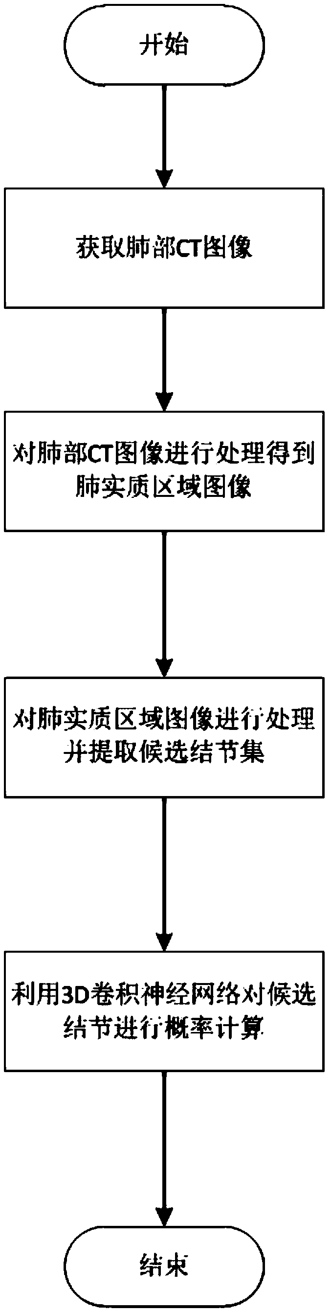
[0071] A method for screening candidate nodules based on CT images, comprising the following steps:

[0072] Step 1. Obtain the lung CT image f to be detected 0 (x,y).

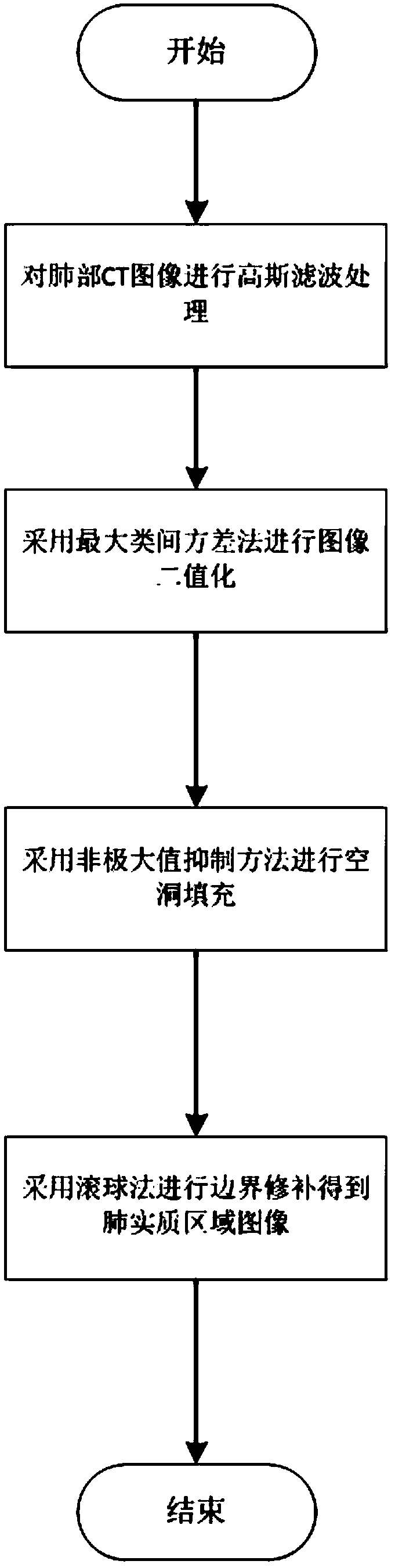
[0073] Step 2. For the lung CT image f in step 1 0 (x, y) perform binarization and extract the lung parenchyma preliminary template f 1 (x,y). Include the following steps:

[0074] Step 2.1. The acquired CT image f to be detected 0 (x, y) is processed by Gaussian filtering to obtain a CT filtered image. Among them, the specific form of the convolution kernel processed by Gaussian filtering is as follows:

[0075]

[0076] Step 2.2. Record the segmentation threshold as T(T∈(0,255)), use the segmentation threshold T as the segmentation value of the CT filter image to segment the foreground image and the background image, record the proportion of the foreground points of the CT filter image to the image as w a1 , the average gray level of the foreground is u a1 , the ratio of the number of background p...


PUM


Abstract

The invention discloses a screening method for candidate nodules based on CT images and relates to the field of medical image processing. The screening method comprises the following steps: step 1, obtaining a to-be-detected lung CT image f0(x, y); step 2, binarizing the lung CT image f0(x, y) in the step 1 and extracting a lung parenchyma preliminary template f1(x, y); step 3, performing lung parenchyma repair and calculation on the lung parenchyma preliminary template f1 (x, y) to obtain a lung parenchyma region image f2 (x, y); step 4, binarizing the lung parenchyma region image f2(x, y) and screening candidate nodules to obtain a candidate nodule set R; step 5, constructing a 3DCNN network structure for classifying candidate nodules; step 6, training the 3DCNN network model according to the 3DCNN network structure and the library lung CT image in a LIDC lung image database; and step 7, determining the probability of each candidate nodule in the candidate nodule set R being a pulmonary nodule according to the 3DCNN network model.

Description

technical field [0001] The invention relates to a method for detecting pulmonary nodules, in particular to a method for screening candidate nodules based on CT images. Background technique [0002] The detection of pulmonary nodules is critical to the processing of lung CT images. CT can provide high-resolution images of the lungs, providing important information for analyzing the presence or absence of pulmonary nodules in lung tissue and the specific location and shape of pulmonary nodules. in accordance with. A huge number of CTs not only provide accurate information, but also greatly increase the workload of radiologists. Therefore, the intelligent processing of CT images came into being. It can not only reduce the labor intensity of doctors and the possible omissions in lung CT images, but also provide accurate quantitative analysis for doctors through image segmentation and feature quantification. [0003] At present, the segmentation of the lung parenchyma in the lu...

Claims


Application Information

Patent Timeline
IPC IPC(8): G06T7/00G06T7/11G06T7/136G06T7/194G06K9/62
CPCG06T7/0012G06T7/11G06T7/136G06T7/194G06T2207/10081G06T2207/20081G06T2207/20084G06T2207/20024G06T2207/30064G06F18/2413G06F18/24147
Inventor 孟明明彭真明蒲红杨吕鹏秦琛烨孙翎马赵学功王卓然曹思颖张天放袁国慧
Owner UNIV OF ELECTRONICS SCI & TECH OF CHINA
Why Eureka
Try Eureka