Cell nucleus segmentation method and system for fluorescence in situ hybridization images

A technology of fluorescence in situ hybridization and cell nucleus, which is applied in the field of cell nucleus segmentation method and system of fluorescence in situ hybridization image, which can solve the problems of blurred nucleus edge and cell adhesion, so as to improve the quality of segmentation, improve over-segmentation, and improve the accuracy rate Effect

Active Publication Date: 2020-11-10
柯凯
View PDF4 Cites 0 Cited by
  • Summary
  • Abstract
  • Description
  • Claims
  • Application Information

AI Technical Summary

Problems solved by technology

[0004] Aiming at the technical problems of fuzzy nuclei in FISH images and difficult identification of cell nuclei and cell adhesion in the images in the prior art, the present invention proposes a cell nucleus segmentation method and system for fluorescence in situ hybridization images

Method used


Image

  • Cell nucleus segmentation method and system for fluorescence in situ hybridization images
  • Cell nucleus segmentation method and system for fluorescence in situ hybridization images
  • Cell nucleus segmentation method and system for fluorescence in situ hybridization images

Examples


Embodiment Construction

[0041] The present invention will be described in further detail below in conjunction with the accompanying drawings and embodiments, but these embodiments should not be construed as limiting the present invention.

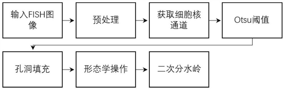
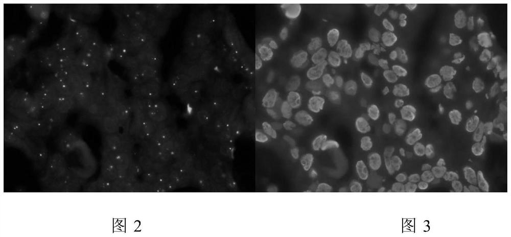
[0042] The present invention proposes a flow process of a cell nucleus segmentation method for fluorescence in situ hybridization images as follows: figure 1 Shown, is for a given FISH image of breast cancer (such as figure 2 shown) to find the process of the nucleus region, the specific steps include:

[0043] 1) The preprocessed FISH image (such as image 3As shown), the pixel value of each pixel of the red, green, and blue three-channel images in the image is reduced by adt_value to ensure that any position of the image is still greater than or equal to 0 after the reduction, and the overall displacement of the image is realized; the value range of adt_value is [2050 ] pixels. Collect the red channel and green channel in the three-channel image where the pi...


PUM


Abstract

The invention discloses a cell nucleus segmentation method and system of fluorescence in situ hybridization images. The method realizes the recognition and segmentation of cell nucleus in FISH imagesthrough pretreatment, binarization, hole filling, morphological operation, secondary watershed and other processing processes. The system comprises a FISH image receiving module, an image preprocessing module, a cell nucleus detecting module and a report generating module. When the FISH images of breast cancer, gastric cancer and other cancer cells are segmented to obtain the cell nucleus region,the invention can reduce the image fluorescence scattering effect through multiple pretreatments, can effectively improve the over-segmentation problem easily caused by the direct watershed, and thusimprove the segmentation quality of the FISH image recognition cell.

Description

technical field [0001] The invention relates to the technical field of medical pathology analysis, which uses a computer image processing method to automatically identify cell nucleus regions in fluorescence in situ hybridization images, and more specifically refers to a method and system for segmenting cell nuclei in fluorescence in situ hybridization images. Background technique [0002] In the field of cancer treatment, such as breast cancer and gastric cancer, the targeted drug Herceptin has the characteristics of high efficiency, low toxicity and strong selectivity. It is a human source of high-purity recombinant DNA derivatives targeting Her-2 / neu receptors Monoclonal Antibodies. Since the targeted drug Herceptin is only effective for breast cancer with HER2 amplification, accurate detection of HER2 amplification is an absolute prerequisite for clinical application of Herceptin, and is also the premise and key to successful targeted therapy. For clinical routine and r...

Claims


Application Information

Patent Timeline
Patent Type & Authority Patents(China)
IPC IPC(8): G06T7/00G06T7/11G06T5/00G06T7/90
CPCG06T5/002G06T5/006G06T5/009G06T7/0012G06T7/11G06T7/90G06T2207/30024
Inventor 刘娟陈哲袁静萍汤永飞贺华段慧芳
Owner 柯凯
Why Eureka
Try Eureka