
A method for image segmentation of pulmonary nodule

A technology for image segmentation and pulmonary nodules, which is applied in the field of computer technology and medical image analysis, can solve problems such as difficult treatment, inability to accurately and automatically segment pulmonary nodules, etc., and achieve the effect of speeding up

Inactive Publication Date: 2018-12-25
UNIVERSITY OF CHINESE ACADEMY OF SCIENCES
View PDF2 Cites 9 Cited by
  • Summary
  • Abstract
  • Description
  • Claims
  • Application Information

AI Technical Summary

Problems solved by technology

[0003] The purpose of the embodiment of the present invention is to provide a pulmonary nodule image segmentation method to solve the problem that the existing medical diagnosis technology cannot accurately and automatically segment pulmonary nodules, which brings difficulties to the treatment

Method used


Image

  • A method for image segmentation of pulmonary nodule
  • A method for image segmentation of pulmonary nodule
  • A method for image segmentation of pulmonary nodule

Examples


Embodiment 1

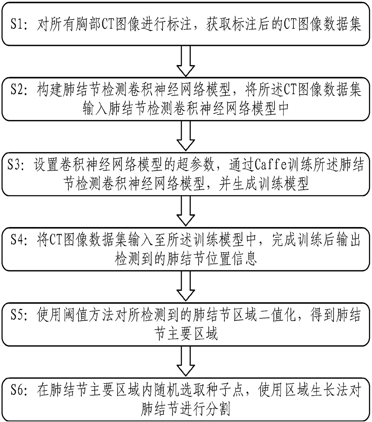
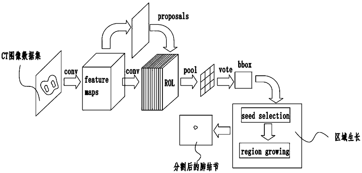
[0033] refer to figure 1 , the present embodiment provides a lung nodule image segmentation method, the pulmonary nodule image segmentation method comprising:

[0034] S1: Label all chest CT images, and obtain the labeled CT image dataset;

[0035] S2: Construct a convolutional neural network model for pulmonary nodule detection, and input the CT image data set into the convolutional neural network model for pulmonary nodule detection;

[0036] S3: Set the hyperparameters of the convolutional neural network model, train the pulmonary nodule detection convolutional neural network model through Caffe, and generate a training model; wherein, the conditions for generating the training model include: when the cost loss function is reduced to an ideal level and When training reaches the required maximum number of iterations.

[0037] S4: Input the CT image data set into the training model, and output the detected pulmonary nodule position information after the training is complete...


PUM


Abstract

The embodiment of the invention discloses a lung nodule image segmentation method, which relates to the fields of computer technology and medical image analysis. The lung nodule image segmentation method comprises the following steps: all chest CT images are annotated to obtain annotated CT image data set; the convolution neural network model of pulmonary nodule detection is constructed and the CTimage data set is input into the convolution neural network model of pulmonary nodule detection; super parameters of the convolution neural network model are set, the convolution neural network modelis trained by Caffe to detect the pulmonary nodules, and a training model is generated; a CT image data set is input into the training model, and the detected pulmonary nodule position information isoutput after completing the training; threshold method is used to binarize the detected pulmonary nodule region, and the main region of pulmonary nodule is obtained. The seed points are randomly selected from the main areas of pulmonary nodules and the nodules are segmented by a region growing method. The invention can solve the problem that the lung nodules can not be accurately and automatically segmented in the prior medical diagnosis technology, and the treatment is difficult.

Description

technical field [0001] The invention relates to the fields of computer technology and medical image analysis, in particular to a lung nodule image segmentation method. Background technique [0002] According to the survey results of the World Health Organization, lung cancer is the cancer with the highest incidence rate and the highest mortality rate. Early detection and treatment of lung cancer is an important measure to reduce lung cancer mortality. With the emergence of medical images, doctors can judge the nature of tumors in time through medical images, so as to provide diagnostic opinions. There are many types of medical images, and among the many types, computed tomography images are most commonly used for lung detection. However, due to the large number of scanned images contained in CT images, it brings a huge workload to doctors. With the continuous development of computer technology, the analysis of medical images by computer can greatly reduce the workload of ...

Claims


Application Information

Patent Timeline
Patent Type & Authority Applications(China)
IPC IPC(8): G06T7/11G06T7/136G06N3/04
CPCG06T7/11G06T7/136G06T2207/10081G06T2207/20081G06T2207/20084G06T2207/30064G06T2207/20104G06N3/045
Inventor 薛健檀彦豪吕科董继阳
Owner UNIVERSITY OF CHINESE ACADEMY OF SCIENCES
Learn More