
Method for conducting MR registration through amplification of bone region and tool design method of MR registration

A regional and bone-based technology, applied in the field of medical imaging, can solve problems affecting the registration accuracy and process, difficult registration process, and low registration accuracy, so as to speed up the registration speed, quickly and smoothly the registration process, and improve The effect of registration accuracy

Inactive Publication Date: 2019-03-08
XIANGYA HOSPITAL CENT SOUTH UNIV
View PDF4 Cites 2 Cited by
  • Summary
  • Abstract
  • Description
  • Claims
  • Application Information

AI Technical Summary

Problems solved by technology

However, in surgery, only a small part of the bone can be exposed through incision. These limited bone areas, as registration mark areas, will greatly affect the accuracy and process of registration. Therefore, the present invention uses 3D printing The fiducial marker tool, which amplifies the smaller bone area into a larger 3D printed registration marker, solves the problems of difficult registration process and low registration accuracy in intraoperative MR

Method used


Image

  • Method for conducting MR registration through amplification of bone region and tool design method of MR registration
  • Method for conducting MR registration through amplification of bone region and tool design method of MR registration
  • Method for conducting MR registration through amplification of bone region and tool design method of MR registration

Examples


Embodiment Construction

[0038] The technical solution of the present invention will be further described in detail below in conjunction with the accompanying drawings and embodiments.

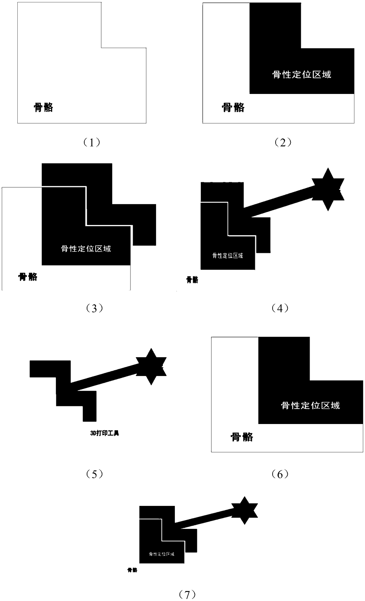
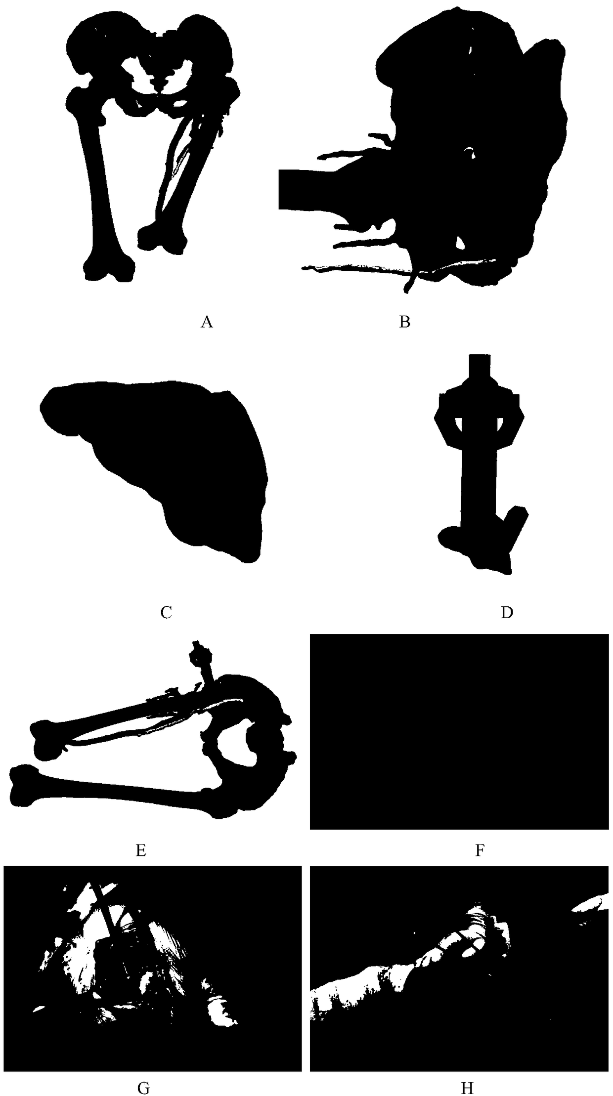
[0039] In the present invention, a certain bone area to be exposed in the surgical area is used as a bony positioning area for three-dimensional reconstruction before operation, and a veneer component is designed according to the bony positioning area, and this component is connected to a reference mark outside the body through a connecting rod , and make this series of parts into a whole tool with 3D printing technology. Before the operation, the data of the patient's bones and surrounding tissues and the digital 3D information of the 3D printed tools are input into the head-mounted (glasses) holographic computer. During the operation, the sterilized 3D printed tool is installed on the corresponding bone positioning area, and then the holographic computer realizes automatic real-time registration by recognizing the f...


PUM


Abstract

The invention belongs to a method of medical treatment imaging, and particularly relates to a method for conducting MR registration through amplification of a bone region and a tool design method of MR registration. Before an operation, an exposed bone region is used as an osseous positioning region for three-dimensional reconstruction, according to the osseous positioning region, an individualized veneer is designed and connected to an extrinsic reference mark through a connecting rod, the series of components are made into a reference mark tool through a 3D printing technology, before the operation, skeleton and peripheral tissue data of a patient and digitalized three-dimensional information of the reference mark tool are input into a holographic computer, in the operation, the sterilized reference mark tool is installed on the corresponding osseous positioning region, a relatively small operation bone region is amplified into a large 3D printing reference mark, the holographic computer realizes registration by recognizing the reference mark on the reference mark tool, in this way, virtual skeletons and other anatomical structures of the patient are overlapped onto the real bodyof the patient, and the operation is easy.

Description

technical field [0001] The invention belongs to medical imaging methods, and in particular relates to a method for expanding a bone region for MR registration and a tool design method thereof. Background technique [0002] Surgical operations require a full understanding of the deep lesions in the human body, but previously they could only be understood through adequate incision exposure, radiography, and Doppler ultrasound. [0003] In recent years, MR technology has provided a new option. The CT and MRI information of the patient's bones and surrounding tissues are input into the head-mounted (glasses) holographic computer through three-dimensional reconstruction. After registration during the operation, the patient can be The virtual three-dimensional image of the bone and surrounding tissue is presented in the patient's body, which fully helps the operator to understand the deep situation of the operation area in real time, regardless of whether the incision is exposed o...

Claims


Application Information

Patent Timeline
Patent Type & Authority Applications(China)
IPC IPC(8): A61B90/00A61B34/10
CPCA61B34/10A61B90/39A61B2034/105A61B2090/3983
Inventor 钟达王成功胡懿郃王小鹏
Owner XIANGYA HOSPITAL CENT SOUTH UNIV
Learn More