Endoscopic image recognition method and device

An image recognition device and image recognition technology are applied in the field of deep learning to achieve the effect of improving inspection quality, reducing workload and reducing pain

Inactive Publication Date: 2019-03-08
青岛美迪康数字工程有限公司
View PDF6 Cites 7 Cited by
  • Summary
  • Abstract
  • Description
  • Claims
  • Application Information

AI Technical Summary

Problems solved by technology

[0004] In order to overcome the above-mentioned deficiencies in the prior art, the purpose of this application is to provide an endoscope image recognition method and device to solve or improve the above-mentioned problems

Method used


Image

  • Endoscopic image recognition method and device
  • Endoscopic image recognition method and device
  • Endoscopic image recognition method and device

Examples


Embodiment Construction

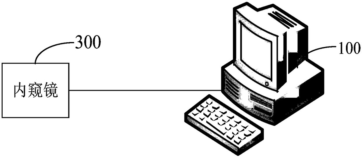
[0037] The following will clearly and completely describe the technical solutions in the embodiments of the present application with reference to the drawings in the embodiments of the present application. Obviously, the described embodiments are part of the embodiments of the present application, not all of them. The components of the embodiments of the application generally described and illustrated in the figures herein may be arranged and designed in a variety of different configurations.

[0038] Accordingly, the following detailed description of the embodiments of the application provided in the accompanying drawings is not intended to limit the scope of the claimed application, but merely represents selected embodiments of the application. All other embodiments obtained by persons of ordinary skill in the art based on the embodiments in the present application without creative efforts shall fall within the protection scope of the present application.

[0039] It should ...


PUM


Abstract

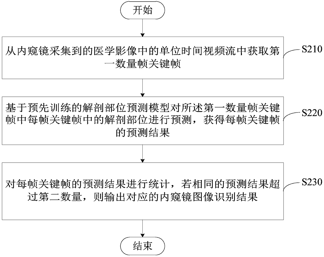
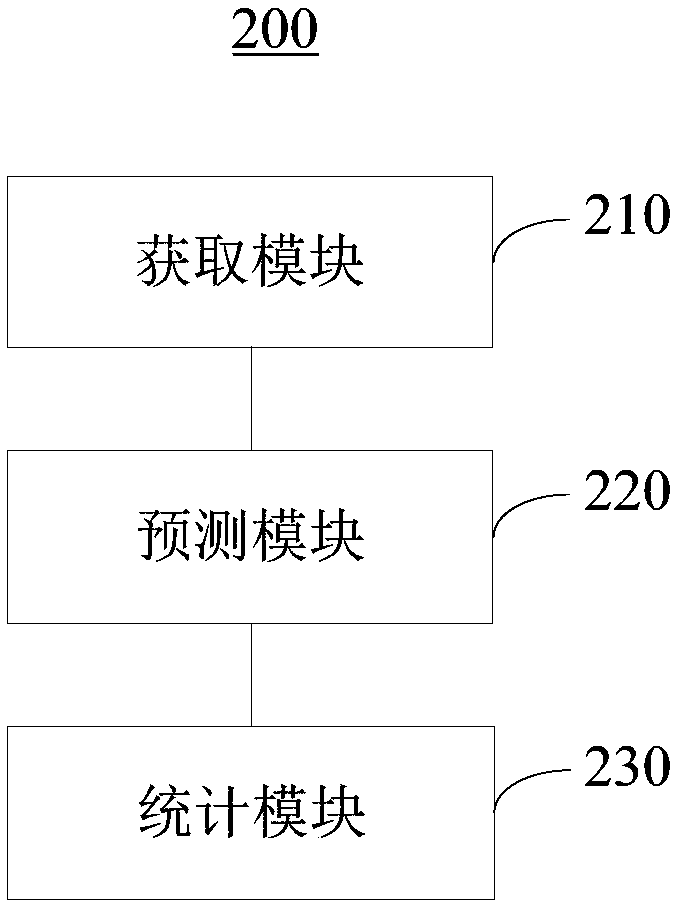
The embodiments of that present application provide the endoscopic image recognition method and device. By acquiring a first number of frame key frames from a unit time video stream in an endoscopically acquired medical image, then, based on the pre-trained anatomical part prediction model, and predicting the anatomical part in each key frame of the first number of frames, a the prediction resultof each key frame is obtained, wherein the prediction result includes the confidence level of each anatomical part in the key frame of the frame. Finally, the prediction result of each key frame is counted, and if the same prediction result exceeds the second number, the corresponding endoscopic image recognition result is output. Thus, the anatomical part in each key frame can be automatically collected and recognized, and the doctor does not need to care about the quantity, quality, and clarity of the collected images, so that the doctor has more energy to focus on the endoscopic operation and observation, thereby lightening the workload of the doctor, improving the examination quality, and reducing the pain of the patient in the examination process.

Description

technical field [0001] The present application relates to the field of deep learning, in particular, to an endoscope image recognition method and device. Background technique [0002] At present, when a doctor performs an endoscopic examination on a patient, each endoscopic examination generally takes several minutes, and the doctor performs dozens or even hundreds of examinations every day. During the examination, the doctor stands next to the endoscope. While operating the endoscope to enter the patient’s body, he also needs to observe and collect images. At the same time, he needs to remember which anatomical parts have been collected and which anatomical parts have not been collected. The energy and physical consumption of the doctor is huge, which increases the intensity of the work, causes the doctor's fatigue, and affects the quality of the examination. [0003] In addition, if the doctor confirms the captured images one by one during the examination, it is necessary...

Claims


Application Information

Patent Timeline
Patent Type & Authority Applications(China)
IPC IPC(8): G06F17/50G06K9/00
CPCG06F30/20G06V20/69
Inventor 邵学军冯健李延青左秀丽李真李广超
Owner 青岛美迪康数字工程有限公司
Why Eureka
Try Eureka