A method for segmenting optic disc based on color retinal fundus image of lesion focus

A fundus image and retina technology, applied in image analysis, fundus mirror, image enhancement and other directions, can solve problems such as poor robustness, and achieve the effect of accurate segmentation

Active Publication Date: 2019-03-08
UNIV OF SHANGHAI FOR SCI & TECH
View PDF6 Cites 4 Cited by
  • Summary
  • Abstract
  • Description
  • Claims
  • Application Information

AI Technical Summary

Problems solved by technology

[0005] The purpose of the present invention is to provide a method for optic disc segmentation based on color retinal fundus images of lesions, to solve the problem of poor robustness of segmentation in the prior art, and to effectively avoid the interference of lesions, blood vessel occlusion, brightness changes, etc., so as to achieve Precise Segmentation of the Optic Disc

Method used


Image

  • A method for segmenting optic disc based on color retinal fundus image of lesion focus
  • A method for segmenting optic disc based on color retinal fundus image of lesion focus
  • A method for segmenting optic disc based on color retinal fundus image of lesion focus

Examples


Embodiment Construction

[0041] The optic disc segmentation method based on the color retinal fundus image of the focus of the present invention will be described in more detail below in conjunction with the schematic diagram, wherein a preferred embodiment of the present invention is shown, it should be understood that those skilled in the art can modify the present invention described here, and still The advantageous effects of the present invention are realized. Therefore, the following description should be understood as the broad knowledge of those skilled in the art, but not as a limitation of the present invention.

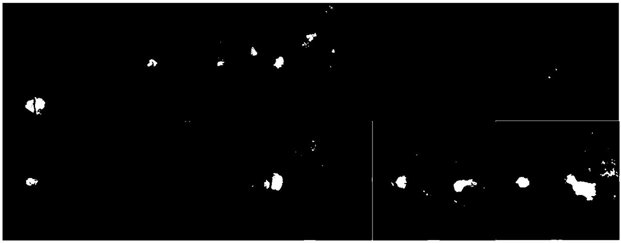
[0042] figure 1 It is part of the images in the Kaggle dataset. These images all have lesions. Many images contain large bright white lesions, and the uneven illumination in the images is also very serious.

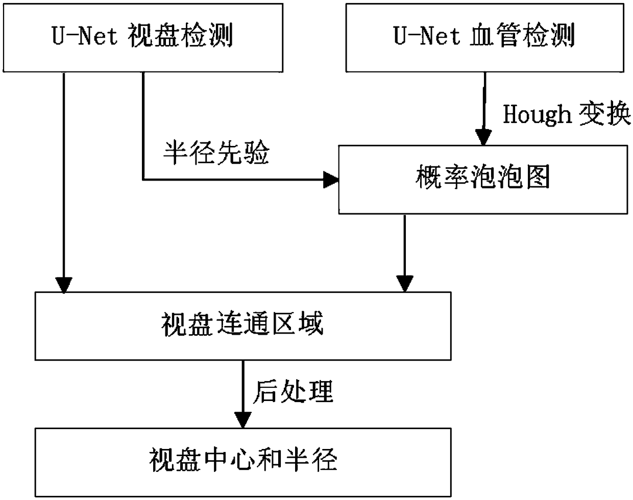
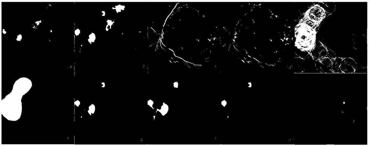
[0043] like figure 2 shown, based on figure 1 The third picture in the first row of the middle table describes the specific content of the technical solution of the presen...


PUM


Abstract

A method for segmenting optic disc based on color retinal fundus image of lesion focus includes the following steps that the blood vessel detection model and optic disc detection model were established, the probabilistic map of blood vessel and the probabilistic map of optic disc were obtained from the detection model of blood vessel and the detection model of optic disc respectively; the probabilistic bubble map of the fitting straight line map of main blood vessel was obtained from the probabilistic map of blood vessel; and the optic disc region was selected from the optic disc communicationarea and the center and radius of optic disc were estimated. The method of the invention can effectively avoid interference of lesions, blood vessel occlusion, brightness change and the like in an image, thereby realizing accurate segmentation of an optic disc.

Description

technical field [0001] The invention belongs to the technical field of detection and segmentation of retinal fundus images, and in particular relates to an optic disc segmentation method based on color retinal fundus images of lesions. Background technique [0002] The automatic analysis of color retinal fundus images has been widely used in the auxiliary diagnosis of various ophthalmology-related systemic diseases such as diabetes, hypertension, kidney disease and neurological diseases. In a normal color fundus image, the macula, fovea, optic disc and vascular network are the four main features that can be observed in the fundus. The optic disc is in the shape of a light yellow or white disc, accounting for about 1% of the area of ​​interest in the entire fundus image. / 5 to 1 / 8, the blood vessels of the fundus diverge from the optic disc area and distribute in the whole fundus in a tree shape. The segmentation of the optic disc plays an extremely important role in the aut...

Claims


Application Information

Patent Timeline
Patent Type & Authority Applications(China)
IPC IPC(8): G06T7/00A61B3/12A61B3/14
CPCA61B3/12A61B3/14G06T7/0012G06T2207/20061G06T2207/20081G06T2207/30041
Inventor 傅迎华李江
Owner UNIV OF SHANGHAI FOR SCI & TECH
Why Eureka
Try Eureka