Brain injury region segmentation method and apparatus for magnetic resonance images acquired on different scanning devices

A nuclear magnetic resonance image and region segmentation technology, applied in image processing and medical fields, can solve problems such as poor lesion segmentation processing effect, and achieve the effect of solving poor processing effect and good lesion segmentation.

Active Publication Date: 2019-03-12
BEIJING SHENRUI BOLIAN TECH CO LTD +1
View PDF5 Cites 0 Cited by
  • Summary
  • Abstract
  • Description
  • Claims
  • Application Information

AI Technical Summary

Problems solved by technology

[0006] The main purpose of the present application is to provide a method and device for brain injury region segmentation for MRI images acquired on different scanning equipment, so as to solve the problem of poor lesion segmentation processing effect on MRI images from different scanning equipment

Method used


Image

  • Brain injury region segmentation method and apparatus for magnetic resonance images acquired on different scanning devices
  • Brain injury region segmentation method and apparatus for magnetic resonance images acquired on different scanning devices
  • Brain injury region segmentation method and apparatus for magnetic resonance images acquired on different scanning devices

Examples


Embodiment Construction

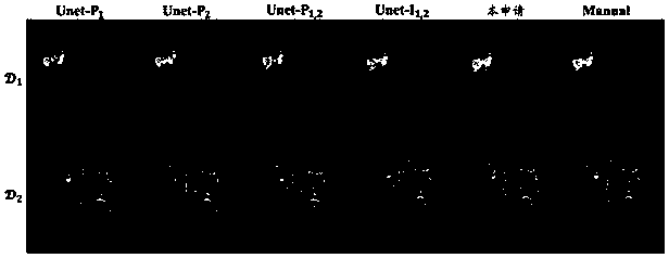
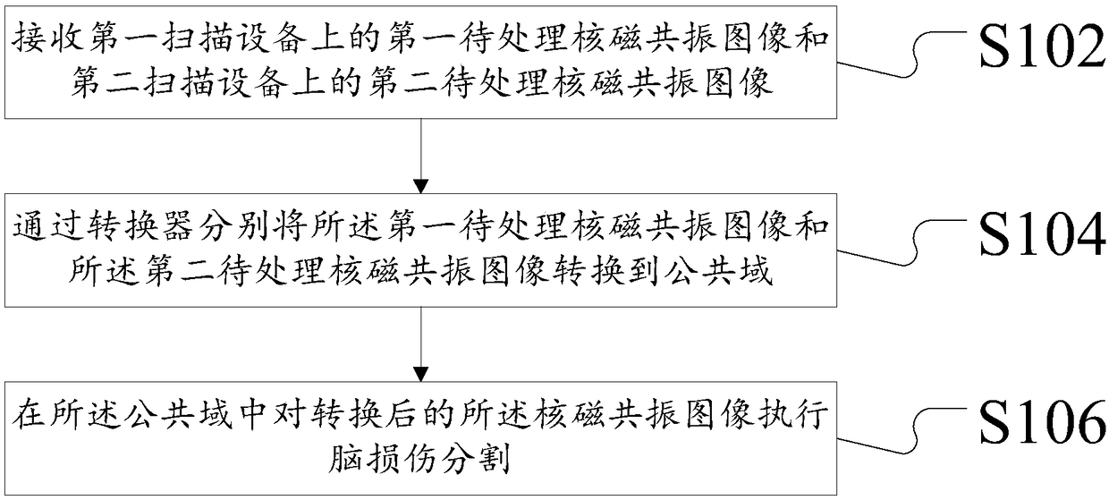
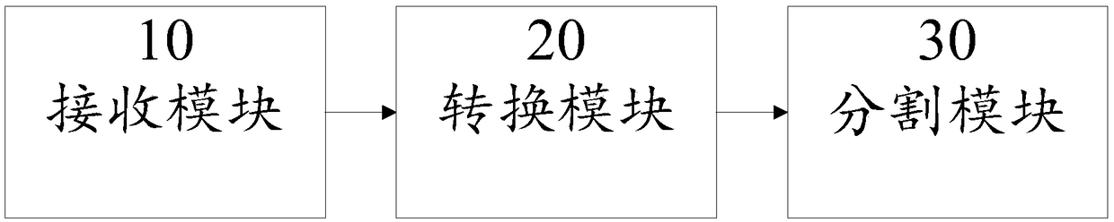
[0028] In order to enable those skilled in the art to better understand the solution of the present application, the technical solution in the embodiment of the application will be clearly and completely described below in conjunction with the accompanying drawings in the embodiment of the application. Obviously, the described embodiment is only It is an embodiment of a part of the application, but not all of the embodiments. Based on the embodiments in this application, all other embodiments obtained by persons of ordinary skill in the art without creative efforts shall fall within the scope of protection of this application.

[0029] It should be noted that the terms "first" and "second" in the description and claims of the present application and the above drawings are used to distinguish similar objects, but not necessarily used to describe a specific sequence or sequence. It should be understood that the data so used may be interchanged under appropriate circumstances for...


PUM


Abstract

The present application discloses a brain injury region segmentation method and apparatus for magnetic resonance images acquired on different scanning devices. The method includes receiving a first nuclear magnetic resonance image to be processed on a first scanning device and a second nuclear magnetic resonance image to be processed on a second scanning device; Converting the first to-be-processed nuclear magnetic resonance image and the second to-be-processed nuclear magnetic resonance image to a common domain by a converter respectively; And performing brain injury segmentation on the converted MRI image in the common domain. The present application solves the technical problem of poor effect of lesion segmentation processing on magnetic resonance images from different scanning apparatuses. The present application can better segment brain lesions on MR images acquired on different scanning devices, thereby accurately detecting cerebral infarction regions.

Description

technical field [0001] The present application relates to the fields of image processing and medical treatment, and in particular, relates to a method and device for segmenting brain injury regions of nuclear magnetic resonance images collected on different scanning devices. Background technique [0002] Cerebral infarction, also known as ischemic stroke, refers to the localized ischemic necrosis or softening of brain tissue caused by cerebral blood supply disturbance, ischemia and hypoxia. Cerebral infarction is the most common type of cerebrovascular disease, accounting for about 75%, with an average fatality rate of 10% to 15%. The disability rate is extremely high, and it is very easy to relapse. The mortality rate of recurrent stroke increases significantly. Therefore, accurate detection of cerebral infarction regions from nuclear magnetic resonance (full name in English: Magnetic Resonance, MR for short) is of great significance for timely detection of risks. [0003]...

Claims


Application Information

Patent Timeline
Patent Type & Authority Applications(China)
IPC IPC(8): G06T7/11G06N3/04
CPCG06T7/11G06T2207/10088G06T2207/30016G06N3/045
Inventor 梁孔明
Owner BEIJING SHENRUI BOLIAN TECH CO LTD
Why Eureka
Try Eureka