Digital pathology system and associated workflow for providing visualized whole-slide image analysis

An image analysis and workflow technology, applied in the field of digital pathology system, can solve the problem that the intuitive visualization does not meet the expected efficiency, and achieve the effect of accurate in vitro diagnosis

Active Publication Date: 2019-05-21
VENTANA MEDICAL SYST INC
View PDF3 Cites 4 Cited by
  • Summary
  • Abstract
  • Description
  • Claims
  • Application Information

AI Technical Summary

Problems solved by technology

[0005] Despite their potential advantages, current digital pathology systems are not as efficient as they can be in providing fast / real-time quantitative analysis of whole tissue sections and their intuitive visualization

Method used


Image

  • Digital pathology system and associated workflow for providing visualized whole-slide image analysis
  • Digital pathology system and associated workflow for providing visualized whole-slide image analysis
  • Digital pathology system and associated workflow for providing visualized whole-slide image analysis

Examples


Embodiment Construction

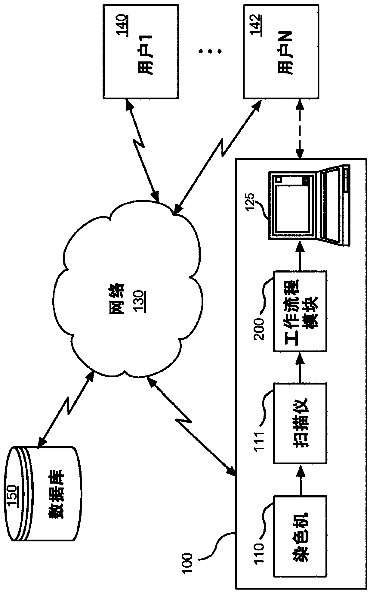
[0039] figure 1 A computer-based digital pathology system 100 is shown operating in a network environment for providing visual quantitative analysis of whole slide images and intuitive visualization of quantification of biomarker expression according to one embodiment of the present invention. The digital pathology system 100 interfaces with a plurality of client computer systems (or user stations) 140 , 142 over a network 130 .

[0040] The digital pathology system 100 may include a stainer 110, a scanner 111, a workflow module 200, a processor or a computer 125, and the like. A user of the client computer system 140, 142, such as a pathologist, histologist, or similar professional, may be able to access, view, and interact with the output of the scanner 111 and workflow module 200 in real time, remotely or locally. . Optionally, these outputs can be stored and accessed on a networked database 150 .

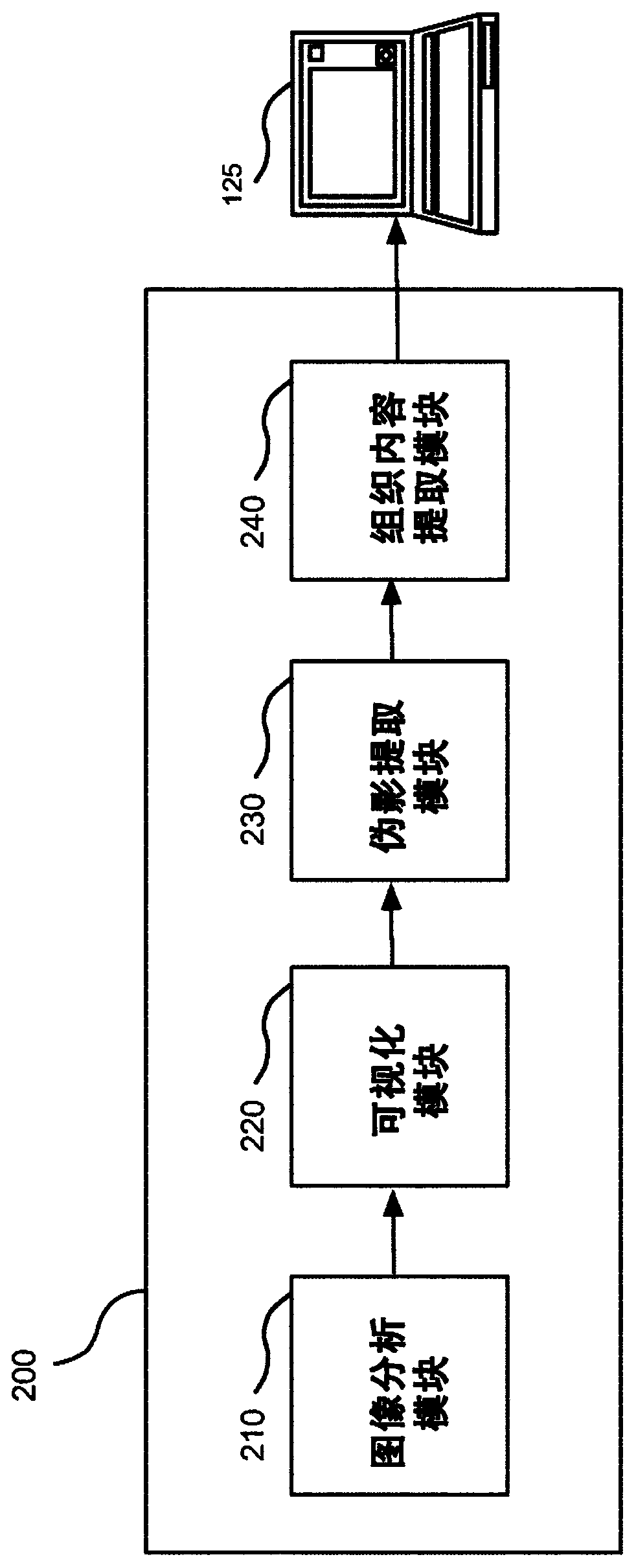
[0041] like figure 2 As further detailed in , the workflow module 200 ...


PUM


Abstract

A digital pathology system and an associated method and computer program product provide a quantitative analysis of entire tissue slides as well as intuitive, effective, fast, and precise quantification of biomarker expressions across relevant areas of the entire tissue slides. The digital pathology system enables a novel workflow that allows the user to efficiently outline clinically relevant morphology in its entirety, including solid tumor areas. Quantitative analysis results are then efficiently and intuitively provided to the user for all tissue content (i.e., millions of cells) within seconds. This efficiency is made possible by a pre-computation step that computes and stores image analysis results for later retrieval. Visualizing vast amount of data effectively is another componentof the system that provides information and confidence to the user about the biomarker expression levels.

Description

technical field [0001] The present invention generally relates to a digital pathology system hosting a workflow for providing quantitative analysis of whole slide images and their intuitive visualization. More specifically, this workflow provides precise quantification of biomarker expression to aid in vitro diagnostics. Background technique [0002] Digital pathology equipment is commonly used to produce digital images of microscope slides. Pathologists and histologists typically visually inspect digital images to obtain information about tissue samples and determine the most appropriate treatment to improve clinical outcomes. [0003] Current digital pathology image analysis systems are increasingly expected to analyze large numbers of high-resolution digital images of tissue sections or "virtual slides". Digital image analysis has proven to be computationally intensive, requires large amounts of storage space, and reduces the ability to perform real-time diagnostics and...

Claims


Application Information

Patent Timeline
Patent Type & Authority Applications(China)
IPC IPC(8): G06T7/00
CPCG06T2207/10056G06T2207/30024G06T2207/30096G06T7/0012G06V20/69G06T7/11G06T2207/30168G06T2207/30204
Inventor M·巴恩斯J·布雷德诺S·楚卡C·居特尔A·洛萨库勒A·萨卡尔E·铃江
Owner VENTANA MEDICAL SYST INC
Why Eureka
Try Eureka