Drainage device for hematocele in ophthalmic vitreous cavity

A vitreous cavity and drainage device technology, which is applied in ophthalmic surgery, laser surgery, etc., can solve the inconvenience and pain of patients

Pending Publication Date: 2019-07-12
厦门浩博尔科技有限公司
View PDF0 Cites 2 Cited by
  • Summary
  • Abstract
  • Description
  • Claims
  • Application Information

AI Technical Summary

Problems solved by technology

At present, for postoperative rebleeding, some of them adopt conservative treatment, such as oral hemostatic drugs, double eye bandaging and immobilization, and some of them adopt secondary operations such as: sitting position gas-liquid exchange, vitreous cavity lavage, vitreous cavity silicone oil filling, etc. Repeated operations bring great harm to patients. great inconvenience and pain

Method used


Image

  • Drainage device for hematocele in ophthalmic vitreous cavity
  • Drainage device for hematocele in ophthalmic vitreous cavity
  • Drainage device for hematocele in ophthalmic vitreous cavity

Examples


Embodiment Construction

[0018] In order to further explain the technical solution of the present invention, the present invention will be described in detail below through specific examples.

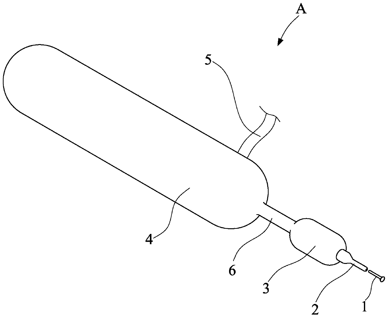
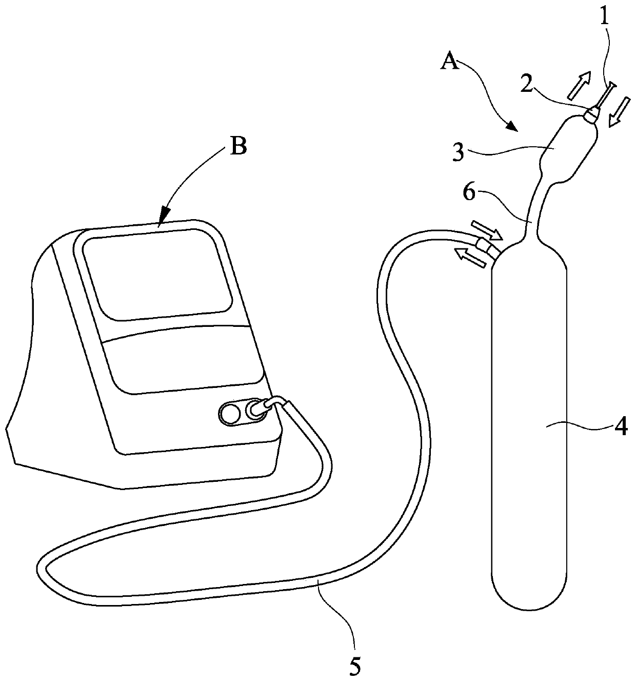
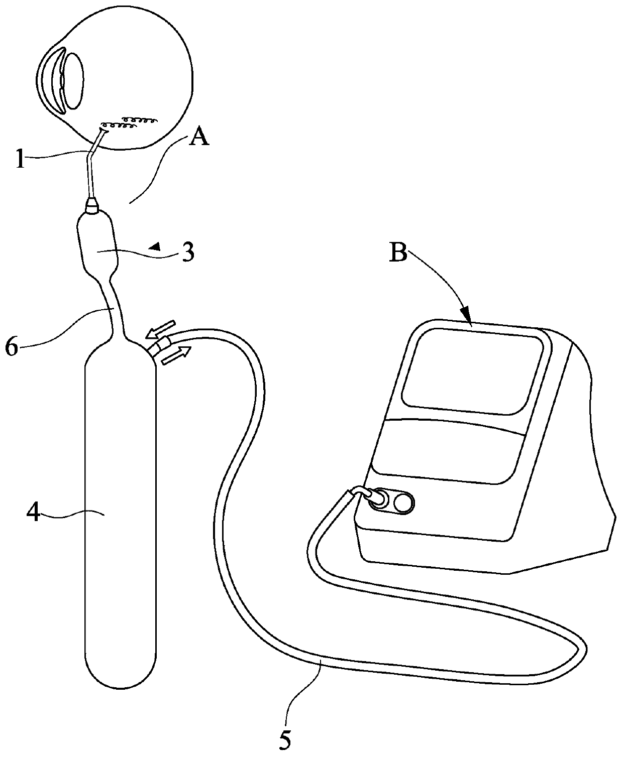
[0019] Such as Figure 1 to Figure 3 As shown, the present invention discloses a drainage device A for ophthalmic vitreous hemorrhage, which includes a disposable drainage tube 1, a catheter 2, a drainage balloon 3, and a rigid capsule 4. The drainage balloon 3 or the rigid capsule 4 One of them is connected with a valve tube 5, and the other end of the valve tube 5 is connected to the host, and the drainage air bag 3 and the rigid bag 4 are communicated through the connecting tube 6, and the drainage tube 1 is inserted on the catheter 2.

[0020] The outer diameter of the drainage tube 1 is 0.4-0.89mm (27-20G), and the inner diameter is 0.3-0.7mm. The head of the drainage tube 1 is made of silica gel or metal, and its shape can be an "L"-shaped hollow pipe structure. It can also be a funnel structure with a w...


PUM


Abstract

The invention discloses a drainage device for hematocele in the ophthalmic vitreous cavity. The drainage device comprises a drainage tube arranged at the head part, a hollow drainage airbag communicated with the drainage tube, and a hollow hard bag communicated with the hollow drainage airbag, and the drainage airbag is arranged between the drainage tube and the hard bag. The drainage tube is connected with the vitreous cavity through a flat part of the ciliary body to achieve the function of collecting the hematocele of the vitreous cavity after a vitrectomy, and the hard bag is arranged outside the body and achieves the function of storing the hematocele; by temporarily squeezing the drainage airbag to increase the intraocular pressure, the function of accelerating liquid drainage to prevent blood clot blockage is achieved, a valve tube can be connected with an external main unit, the intraocular pressure is monitored in real time, the pressure is adjusted through the main unit, andsafe gas is input or output to achieve the purpose of stabilizing the intraocular pressure. The hematocele is drained into the hard bag, whether or not the drainage device is detached is determined according to the judgment about whether the drainage liquid is clear or not, and by directly draining the hematocele outside the body, the situation is avoided that great inconvenience and pain are brought to patients due to repeated hematocele secondary operations.

Description

technical field [0001] The invention relates to an auxiliary treatment device after ophthalmic clinical vitrectomy, in particular to a drainage device for ophthalmic vitreous cavity hemorrhage. Background technique [0002] Vitreous hemorrhage is a common complication of visual hazard from ocular trauma or retinal or choroidal vascular disease. Hemorrhage not only makes the refractive medium cloudy, but also can cause serious damage to the eye tissue; in different cases, the consequences of vitreous cavity hemorrhage are very different, in fundus hemorrhage-related diseases such as diabetic retinopathy, choroidal polypoid lesions, retinal During vitrectomy for venous occlusion, traumatic vitreous hemorrhage, wet age-related macular degeneration, etc., repeated postoperative bleeding is easy. Repeated bleeding not only affects vision, but also brings great harm to patients, such as corneal blood staining, ghost cell glaucoma, proliferative retinopathy, tractive retinal detac...

Claims


Application Information

Patent Timeline
Patent Type & Authority Applications(China)
IPC IPC(8): A61F9/007
CPCA61F9/00736A61F9/00781A61F2009/00874
Inventor 李海波
Owner 厦门浩博尔科技有限公司
Why Eureka
Try Eureka