Cell detection segmentation system and method based on a deep learning neural network

A neural network and deep learning technology, applied in the field of cell detection and segmentation systems, can solve problems such as taking a long time and effort, and achieve the effect of simple implementation and less hardware

Pending Publication Date: 2019-07-26
深圳市麦迪普科技有限公司
View PDF6 Cites 13 Cited by
  • Summary
  • Abstract
  • Description
  • Claims
  • Application Information

AI Technical Summary

Problems solved by technology

However, pathological images are usually super-large pixel images. The traditional method r

Method used


Image

  • Cell detection segmentation system and method based on a deep learning neural network
  • Cell detection segmentation system and method based on a deep learning neural network
  • Cell detection segmentation system and method based on a deep learning neural network

Examples


Embodiment Construction

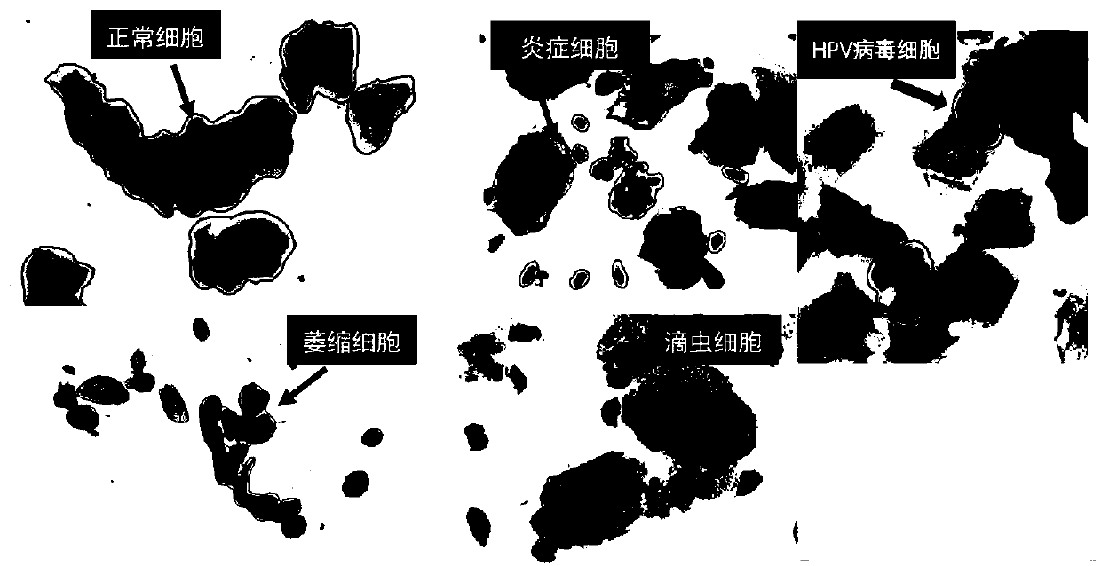
[0052]The embodiment of the present invention provides a cell detection and segmentation system and method based on deep learning neural network, which is used to detect cytopathological images, and can detect normal cells, inflammatory cells, trichomonas cells, atrophic cells and HPV virus in the picture After the cells are separated, mark each type and count their number, analyze whether the patient is infected, inflamed, etc., and provide a reliable and efficient auxiliary diagnosis basis for pathologists. Example pictures of cell types such as figure 1 shown. In order to enable those skilled in the art to better understand the solutions of the present invention, the technical solutions in the embodiments of the present invention will be clearly and completely described below in conjunction with the drawings in the embodiments of the present invention. Based on the embodiments of the present invention, all other embodiments obtained by persons of ordinary skill in the art ...


PUM


Abstract

The invention provides a cell detection segmentation system and method based on a deep learning neural network. A deep learning method is used for detecting a cell pathological image. Normal cells, inflammatory cells, trichomonad cells, atrophy cells, HPV viruses and other cells in the picture are segmented out. Each type is marked and the number of each type is counted. A judgment is made as to whether a patient is infected or inflammatory or not and reliable and efficient auxiliary diagnosis is provided for pathologists. The invention has the advantages of simplicity, effectiveness, less required hardware configuration and low implementation cost.

Description

technical field [0001] The present invention relates to the application of deep learning technology in medical image processing, in particular to a system and method for cell detection and segmentation based on deep learning neural network. Background technique [0002] Cervical cancer is an important killer of women's health. Its incidence rate ranks second among female malignant tumors, second only to breast cancer. Statistics show that about 20,000 to 30,000 people die from cervical cancer every year, and it is on the rise and younger. TCT cervical anti-cancer examination is an important examination method, which can detect cervical cancer cells, as well as some precancerous lesions and microbial infections such as mold, trichomonas, viruses, and chlamydia. However, pathological images are usually super-large pixel images. Traditional methods require pathologists to check the entire film, which takes a long time and a lot of energy. Deep learning technology has develop...

Claims


Application Information

Patent Timeline
IPC IPC(8): G06T7/00G06T7/11
CPCG06T7/0012G06T7/11G06T2207/30024G06T2207/20104G06T2207/20081Y02A90/10
Inventor 沈琳琳谢鑫鹏蔡盛灶
Owner 深圳市麦迪普科技有限公司
Why Eureka
Try Eureka