
Imaging device for cone cells

An imaging device and cone cell technology, applied in medical science, ophthalmoscope, equipment for testing eyes, etc., can solve problems affecting image quality

Pending Publication Date: 2019-08-02
广东唯仁医疗科技有限公司
View PDF2 Cites 1 Cited by
  • Summary
  • Abstract
  • Description
  • Claims
  • Application Information

AI Technical Summary

Problems solved by technology

The human eye itself is an optical system, and the optical system often has various aberrations in the process of measuring the human eye, which seriously affects the image quality

Method used


Image

  • Imaging device for cone cells
  • Imaging device for cone cells

Examples


Embodiment Construction

[0022] The idea, specific structure and technical effects of the present invention will be clearly and completely described below in conjunction with the embodiments and accompanying drawings, so as to fully understand the purpose, scheme and effect of the present invention. It should be noted that, in the case of no conflict, the embodiments in the present application and the features in the embodiments can be combined with each other.

[0023] It should be noted that, unless otherwise specified, when a feature is called "fixed" or "connected" to another feature, it can be directly fixed and connected to another feature, or indirectly fixed and connected to another feature. on a feature. In addition, descriptions such as up, down, left, and right used in the present invention are only relative to the mutual positional relationship of the components of the present invention in the drawings. As used herein and in the appended claims, the singular forms "a", "the", and "the" ar...


PUM


Abstract

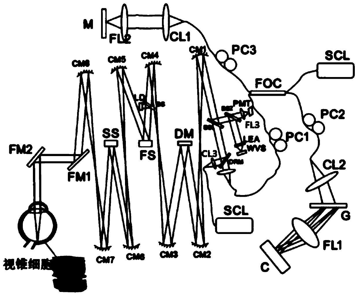
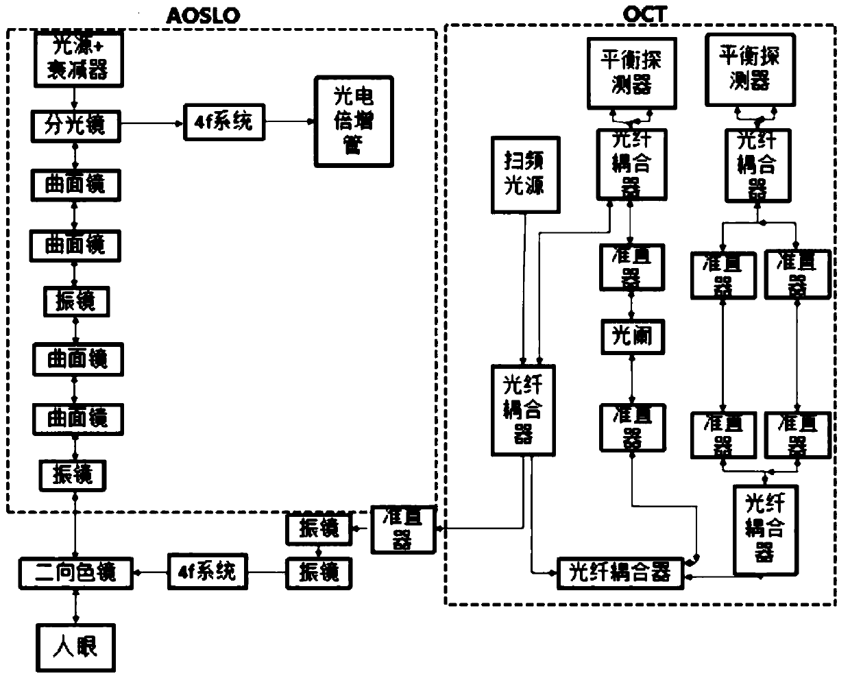
The invention provides an imaging device for cone cells. The imaging device comprises an AOSLO (Adaptive Optical Scanning Laser Ophthalmoscope), an OCT (Optical Coherence Tomography) and an imaging processing device. A light beam entering into cone cells of fundus is processed and returned through OCT and then is subjected to interference treatment with the light beam processed by AOSLO, and a processed interference signal is sent to the imaging processing device for generating a high-precision three-dimensional image, so that the activity of the cone cells of fundus can be observed.

Description

technical field [0001] The present invention generally relates to an imaging device, in particular to a cone cell imaging device. Background technique [0002] The application of adaptive optics (AO) in ophthalmology is to adaptively correct various aberrations of the optical system through the wavefront sensor-wavefront controller, and greatly improve the lateral resolution of the system. The human eye itself is an optical system, and the optical system often has various aberrations in the process of measuring the human eye, which seriously affects the image quality. Introducing adaptive optics technology into the optical system can solve this problem. This patent combines Adaptive Optics Scanning Laser Ophthalmoscope (AOSLO) with Optical Coherence Tomography (OCT) to greatly improve the resolution of the system in two-dimensional space and observe the cone-level activities caused by the system. The activity of the retinal layers can assess the activity of cone cells. ...

Claims


Application Information

Patent Timeline
Patent Type & Authority Applications(China)
IPC IPC(8): A61B3/12A61B3/14
CPCA61B3/102A61B3/12A61B3/14
Inventor 邓卓健吴小翠
Owner 广东唯仁医疗科技有限公司
Learn More