
Tumor pathological image display station

A pathological image and display platform technology, which is applied in the field of tumor pathological image display platform, to achieve the effect of simple operation, good display effect, and convenient observation and analysis of image details

Active Publication Date: 2019-08-27
HENAN PROVINCE HOSPITAL OF TCM THE SECOND AFFILIATED HOSPITAL OF HENAN UNIV OF TCM
View PDF13 Cites 13 Cited by
  • Summary
  • Abstract
  • Description
  • Claims
  • Application Information

AI Technical Summary

Problems solved by technology

[0004] In view of the above problems, the present invention provides a tumor pathology image display table, which solves the problems mentioned in the background technology

Method used

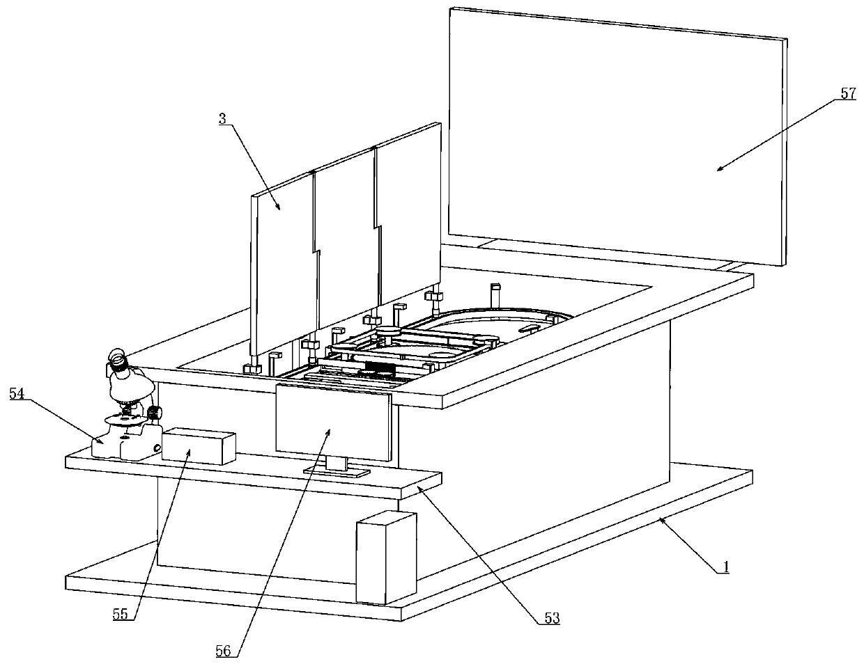

Image

  • Tumor pathological image display station
  • Tumor pathological image display station
  • Tumor pathological image display station

Examples


Embodiment 1

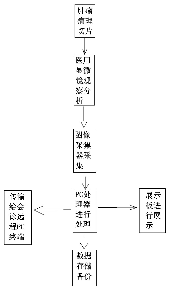
[0036] Embodiment 1, in conjunction with the attached Figure 1-17 , a tumor pathological image display platform, including a support 1, the support 1 is connected with an operation table 53, the upper end of the support 1 is vertically arranged with side walls, and the operation table 53 is arranged on a side wall, so The operating table 53 is provided with a medical microscope 54, the medical microscope 54 is electrically connected to an image collector 55, and the image collector 55 is electrically connected to a PC processor 56, and the processed tumor pathological slices are placed in the image collector 56. Observe under the medical microscope 54, collect the controversial pathological images under the medical microscope 54 using the image collector 55, and then transmit the data to the PC processor 56 for processing to obtain an electronic version of the tumor pathological slice image, and then we can Print it. If remote consultation is required, transfer the image data...

Embodiment 2

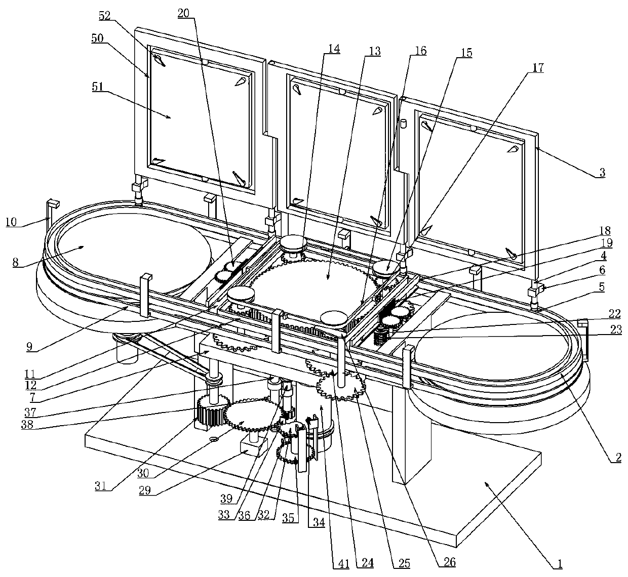
[0040] Embodiment 2, on the basis of Embodiment 1, combined with the attached Figure 1-17, the drive device includes a drive motor 29 installed on the support 1, the drive motor 29 is connected to the controller, the drive motor 29 is a forward and reverse rotation motor, and is controlled and driven by the controller through the trigger of the limit switch 27 The rotation direction of the motor 29, the output shaft of the drive motor 29 is coaxially connected to the fifth gear 30, and the fifth gear 30 meshes with a first one-way gear 31 rotatably connected to the lower end of the bracket 7. The first one-way The structure of the gear 31 is that a one-way bearing is fixed on the rotating shaft, and the one-way bearing is sleeved with a natural spur gear to ensure that the first one-way gear 31 only transmits power in one direction. One of the main pulleys 8 is connected through a belt to provide power input for the drive belt 9. The fifth gear 30 meshes with a second incompl...

Embodiment 3

[0041] Embodiment 3, on the basis of Embodiment 2, combined with the attached Figure 1-17 , the lifting device includes a quill 41 rotatably connected to the upper end of the support 1 and a lead screw 42 rotatably connected to the lower end of the bracket 7, the lead screw 42 is coaxially threadedly sleeved in the quill 41, and the The sleeve shaft 41 is connected with the rotating shaft of the seventh gear 36 through a belt. The lifting device here is realized by the sleeve-screw 42 structure. This structure has a good self-locking function, and the transmission is stable and reliable. .


PUM


Abstract

The invention relates to a tumor pathological image display station. Through a rectangular track formed by a set of elliptical tracks and longitudinal tracks connected therebetween, display boards canbe freely switched between the elliptical tracks and the longitudinal tracks, and the lower ends of the display boards are provided with pushing plates; the positions of driving rods on a driving belt and the positions of pushing rods on a guide belt in the vertical direction are changed, so as to drive the pushing plates to drive the display boards to be switched between the elliptical tracks and rectangular track respectively; a large fluted disc is meshed with two sets of first incomplete gears, a second incomplete gear is meshed with a rack through a second gear, and the elliptical tracksand the rectangular track are separated by the rack; a driving device provides power input and reversing action, the structure is clever, the operation is simple, the elliptical track show images tomore people, facilitating observation and analyzing of the details of the images, the rectangular track allows repeated flip of the display boards, facilitating comparative analysis; the display effect is good, and the accuracy of medical staff for tumor pathological diagnosis is improved.

Description

technical field [0001] The invention belongs to the technical field of display tables, and particularly relates to a tumor pathological image display table. Background technique [0002] Tumors refer to new organisms formed by the proliferation of local tissue cells under the action of various tumorigenic factors, because these new organisms are mostly space-occupying massive protrusions, also known as neoplasms. According to the cell characteristics of new organisms and the degree of harm to the body, tumors are divided into two categories: benign tumors and malignant tumors, and cancer is the general term for malignant tumors. Cancer patients have a very low chance of being cured. Therefore, as a natural enemy of human beings, cancer, as a natural enemy, should be diagnosed and treated as soon as possible, and the lesions should be strangled in swaddling clothes. [0003] The diagnosis of tumor pathology is an important means of diagnosing tumors and plays a crucial role....

Claims


Application Information

Patent Timeline
Patent Type & Authority Applications(China)
IPC IPC(8): A61B5/00
CPCA61B5/00
Inventor 刘粉霞雷彦宏
Owner HENAN PROVINCE HOSPITAL OF TCM THE SECOND AFFILIATED HOSPITAL OF HENAN UNIV OF TCM
Learn More