A kind of functional modification method of cell membrane
A modification method and cell membrane technology, applied in the field of cell engineering, can solve problems such as poor stability, shedding, and reduced ability of probe target recognition.
- Summary
- Abstract
- Description
- Claims
- Application Information
AI Technical Summary
Problems solved by technology
Method used
Image
Examples
Embodiment 1
[0077] Embodiment 1: The stability of the amphiphilic tetrahedral probe of the present invention on the cell membrane
[0078] Reduced shedding rate of amphiphilic tetrahedral probes
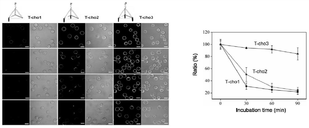
[0079] After cell counting, 200,000 CEM cells were taken, washed once with PBS, and equally divided into 2 groups, and then mixed with 250 nM S-cho1-FAM (sequence: CCCAGGTTCTCTT- / i6FAMdT / -TTTTTTTTTTTTTT-cholesterol, i6FAMdT represents the place The base T is modified by a fluorescent group) and 250nM T-cho3-FAM prepared by the method of the present invention were co-incubated at 4°C for 10 minutes, washed once with PBS, and dispersed into 1640 medium containing 10% FBS. The two groups of cells were cultured at 37°C in a 5% carbon dioxide environment for 0 hour, 0.5 hour, 1 hour, and 1.5 hour, washed once with PBS, and confocal imaging was used to observe the probe shedding. Such as Figure 6 As shown, the probe S-cho1-FAM was almost completely shed within half an hour, while the probe T-cho3-F...
Embodiment 2
[0080] Example 2: The reduced endocytic efficiency of the amphiphilic tetrahedral probe of the present invention
[0081] After cell counting, 200,000 CEM cells were taken, washed once with PBS, and evenly divided into 2 groups, and then co-incubated with 250nM S-cho1-FAM and 250nM T-cho3-FAM at 4°C for 10 minutes, PBS Wash once and disperse in PBS containing 5 mM magnesium chloride. Both groups of cells were incubated at 37°C for 15 minutes, and the endocytosis of the probe was observed by confocal imaging. Such as Figure 7 As shown, the probe S-cho1-FAM was obviously endocytized after being cultured for 15 minutes, but the probe T-cho3-FAM had no obvious endocytosis. It is revealed that the amphiphilic tetrahedral probe greatly improves the stability on the cell membrane compared with the traditional amphiphilic probe.
Embodiment 3
[0082] Example 3: The amphiphilic tetrahedron probe of the present invention enhances the target recognition ability on the cell membrane
[0083] After cell counting, 200,000 Ramos cells were collected and stained with red live cell tracer (CellTracker TM) stained at 37°C for 15 minutes, washed once with PBS, and divided into 2 groups. Then co-incubated with 250nM T-cho3-sgc8 probe and 250nM S-cho1-sgc8 probe at 4°C for 10 minutes, washed once with PBS, and dispersed into DPBS containing 2% bovine serum albumin (BSA) . Take another 2 million CEM cells, divide them into two groups, add them to the above two groups of Ramos cells, and shake (240rpm) at room temperature for 10 minutes, 20 minutes, 30 minutes, 60 minutes, 90 minutes, 120 minutes, 240 minutes, 360 minutes, 540 minutes. The samples at different time points were imaged with a confocal microscope, and the aggregation efficiency of the cell map was counted. Such as Figure 8 As shown, the T-cho3-sgc8 probe-modifi...
PUM
 Abstract
Description
Claims
Application Information
 - R&D
- Intellectual Property
- Life Sciences
- Materials
- Tech Scout
- Unparalleled Data Quality
- Higher Quality Content
- 60% Fewer Hallucinations
Browse by: Latest US Patents, China's latest patents, Technical Efficacy Thesaurus, Application Domain, Technology Topic, Popular Technical Reports.
© 2025 PatSnap. All rights reserved.Legal|Privacy policy|Modern Slavery Act Transparency Statement|Sitemap|About US| Contact US: help@patsnap.com


