Method and system for automatically segmenting esophagus cancer radiotherapy target area and organs at risk

An automatic segmentation, esophageal cancer technology, applied in the fields of instruments, image analysis, character and pattern recognition, etc., can solve the problems of poor segmentation effect, sensitive model quality, and the segmentation effect is difficult to meet clinical requirements, etc., to achieve great clinical application potential, The effect of low missed or false detection rate and improved accuracy

Inactive Publication Date: 2019-09-06
ANHUI UNIVERSITY
View PDF5 Cites 10 Cited by
  • Summary
  • Abstract
  • Description
  • Claims
  • Application Information

AI Technical Summary

Problems solved by technology

[0003] Existing technology 1 uses a region segmentation algorithm based on region features. The basic idea of ​​the algorithm is to start from a group of growing points, which can be a single pixel or a small region. Neighboring pixels or regions are merged with growing points to form new growing points, and this process is repeated until it cannot grow; this algorithm is not effective for the segmentation of regions with little difference in gray distribution
[0004] The second prior art uses a medical image segmentation algorithm based on a deformation model. This algorithm first gives a closed curve corresponding to the approximate position of the object boundary on the image, that is, the initial model; then the curve moves under the guidance of the image information and the information of the curve itself. That is, the deformation process of the model; the final curve moves to the correct object boundary and stops, that is, the model converges; the algorithm is sensitive to the quality of the model, and the segmentation effect is difficult to meet the clinical requirements

Method used


Image

  • Method and system for automatically segmenting esophagus cancer radiotherapy target area and organs at risk
  • Method and system for automatically segmenting esophagus cancer radiotherapy target area and organs at risk
  • Method and system for automatically segmenting esophagus cancer radiotherapy target area and organs at risk

Examples


Embodiment Construction

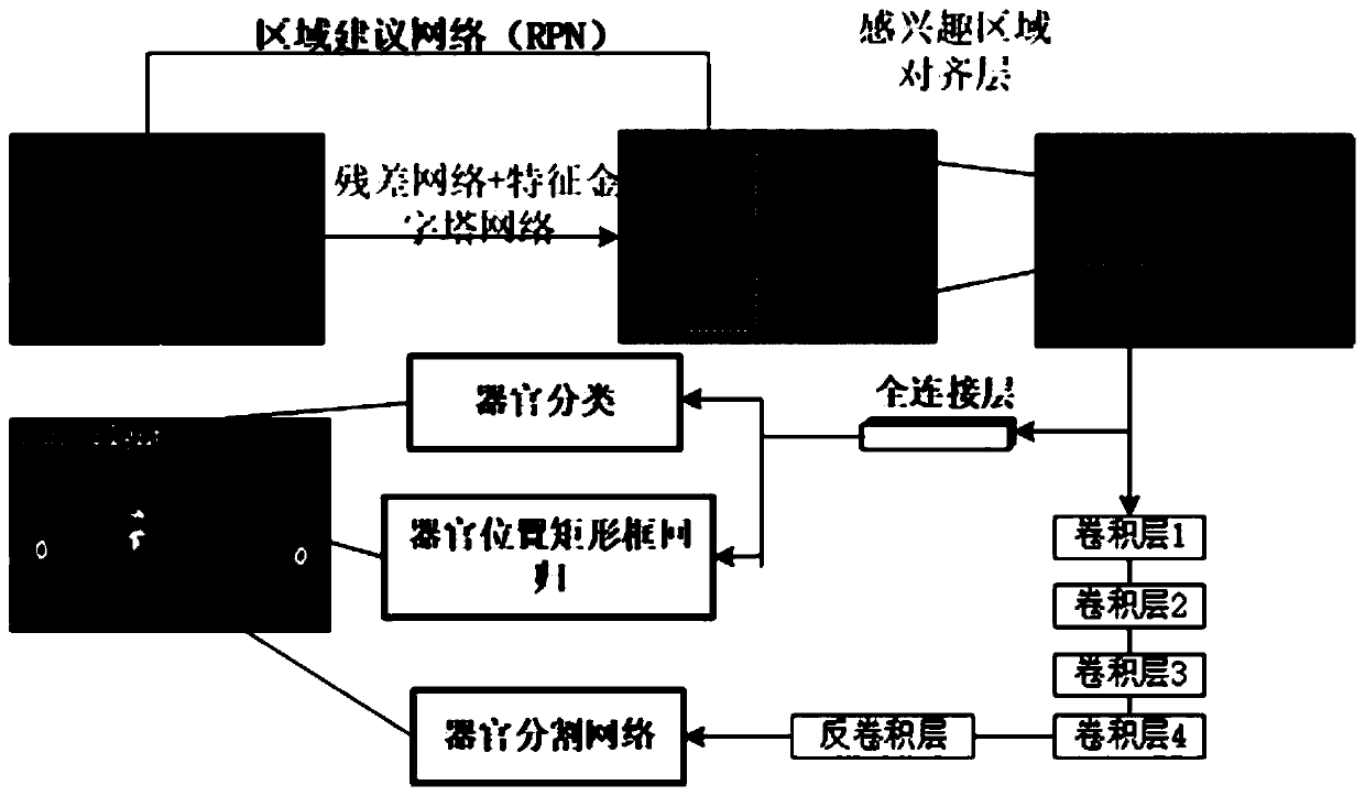
[0037] The method for automatically segmenting esophageal cancer radiotherapy targets and organs at risk in the present invention uses the Mask R-CNN algorithm. The Mask R-CNN algorithm is an instance segmentation algorithm that can be used for target detection, target instance segmentation, and target key point detection.

[0038] Such as figure 1 As shown, the present invention automatically divides the method for esophageal cancer radiotherapy target volume and endangered organs, comprises the following steps:

[0039] Step A, extract the texture, color and other features of the input CT image through the residual network (ResNet), fuse the multi-scale feature map through the feature pyramid network (FPN), and use the region proposal network (RPN) to analyze each point in the feature map. region of interest (ROI) for screening.

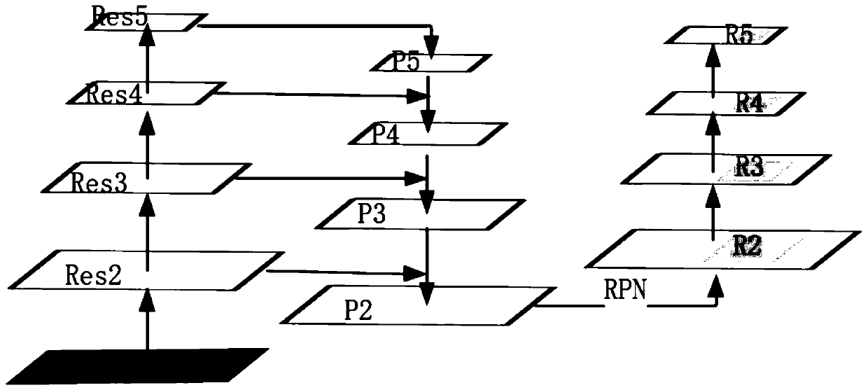
[0040] Such as figure 2 As shown, the specific steps of step A are as follows:

[0041] Step a1, input a medical CT image containing multiple ...


PUM


Abstract

The invention discloses a method and a system for automatically segmenting an esophageal cancer radiotherapy target area and organs at risk, and relates to the field of medical image segmentation. Themethod comprises the following steps: extracting features of an input CT image through a residual network, and fusing multi-scale feature maps through a feature pyramid network; screening the regionof interest of each point in the feature map through a region suggestion network; pooling the region-of-interest screened by the region suggestion network to a fixed size in combination with the region-of-interest alignment layer; inputting a region of interest pooled to a fixed size into the full connection layer for organ classification, and performing organ position border regression; meanwhile, inputting the interest area pooled to a fixed size into the organ segmentation network. The method has the advantage that the accuracy of automatically segmenting the esophagus cancer radiotherapy target area and various organs at risk is improved.

Description

technical field [0001] The invention relates to the field of medical image segmentation, in particular to a method and system for automatically segmenting radiotherapy target areas and endangered organs of esophageal cancer. Background technique [0002] Esophageal cancer is a primary malignant tumor of the esophagus, affecting as many as 200,000 people each year in China. Radiation therapy is one of the main treatment methods for esophageal cancer. Its treatment planning is highly dependent on the accurate segmentation of the planning target volume PTV and multiple organs at risk OAR. The accuracy of segmentation determines the quality of radiation therapy planning dose optimization, which directly affects The success or failure of radiotherapy or the incidence of complications. Due to the variable shape of the target area of ​​esophageal cancer and the differences between human bodies, the boundary between the target area and organs is blurred. In clinical practice, docto...

Claims


Application Information

Patent Timeline
Patent Type & Authority Applications(China)
IPC IPC(8): G06T7/11G06K9/32G06K9/62
CPCG06T7/11G06T2207/10081G06T2207/20016G06T2207/20084G06T2207/20081G06T2207/30061G06V10/25G06V2201/03G06F18/241
Inventor 李腾刘剑飞王妍于明晖
Owner ANHUI UNIVERSITY
Why Eureka
Try Eureka