Micro calcification point automatic detection method based on ultrasonic breast tumor image

A technology for automatic detection of breast tumors, applied in image enhancement, image analysis, image data processing, etc., can solve problems such as difficulty in algorithm implementation

Active Publication Date: 2019-09-20
南京天智信科技有限公司
View PDF15 Cites 4 Cited by
  • Summary
  • Abstract
  • Description
  • Claims
  • Application Information

AI Technical Summary

Problems solved by technology

[0006] The purpose of the present invention is to overcome the problem that the existing low-contrast ultrasonic mammary gland images are used to detect tiny calcifications, and the algorithm is difficult to implement, so as to assist doctors in diagnosing calcific

Method used


Image

  • Micro calcification point automatic detection method based on ultrasonic breast tumor image
  • Micro calcification point automatic detection method based on ultrasonic breast tumor image
  • Micro calcification point automatic detection method based on ultrasonic breast tumor image

Examples


Embodiment Construction

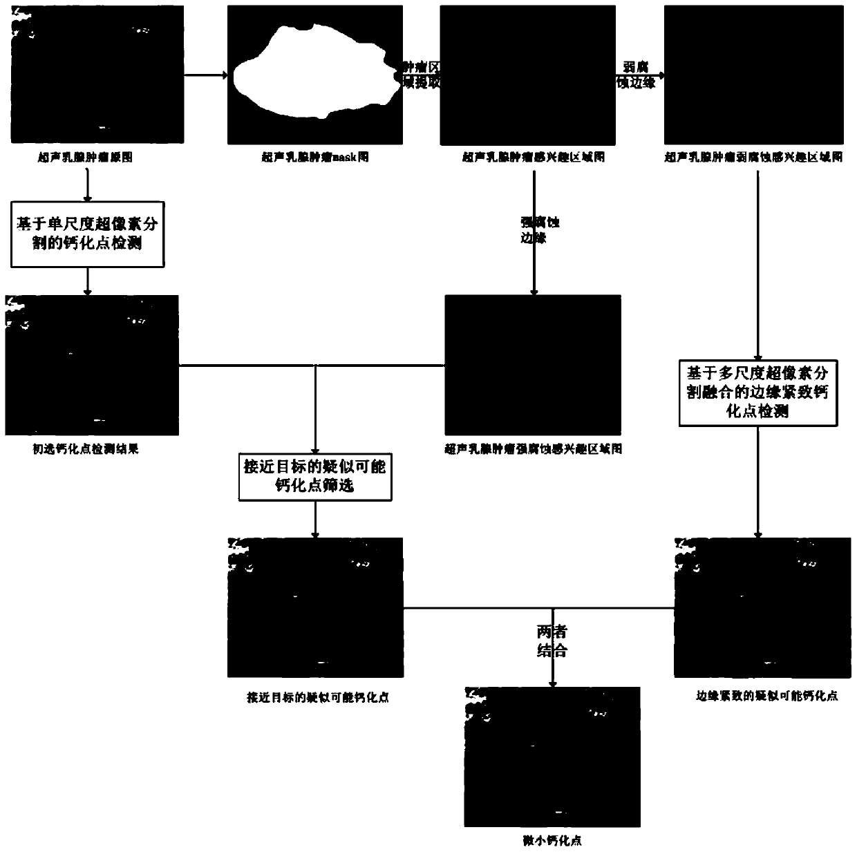
[0070] The present invention will be further described below in conjunction with the accompanying drawings.

[0071] Such as figure 1 As shown, the automatic detection method of tiny calcification points based on ultrasound breast tumor images of the present invention comprises the following steps,

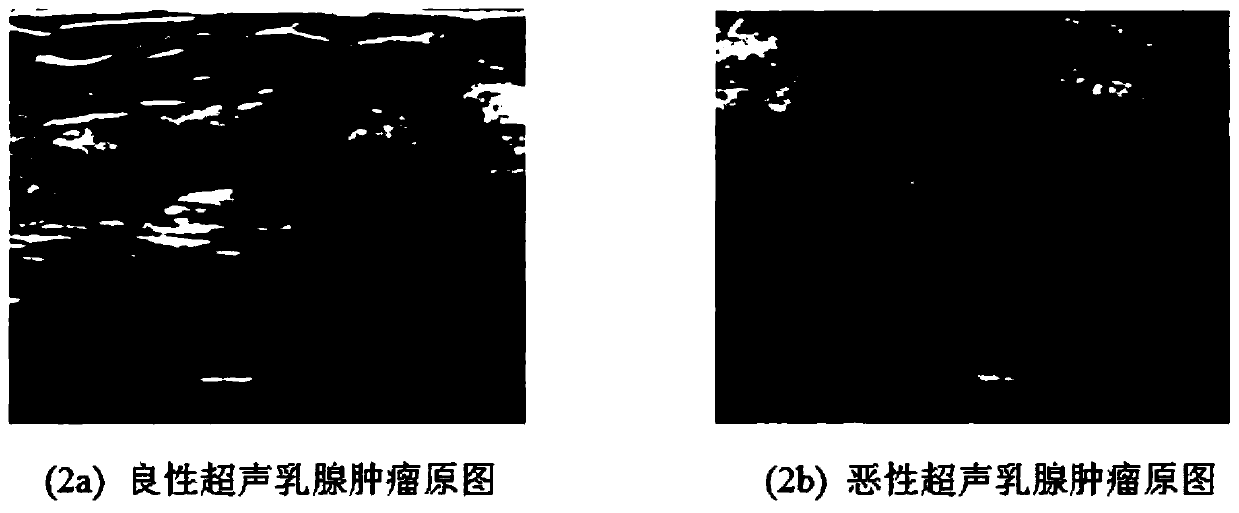
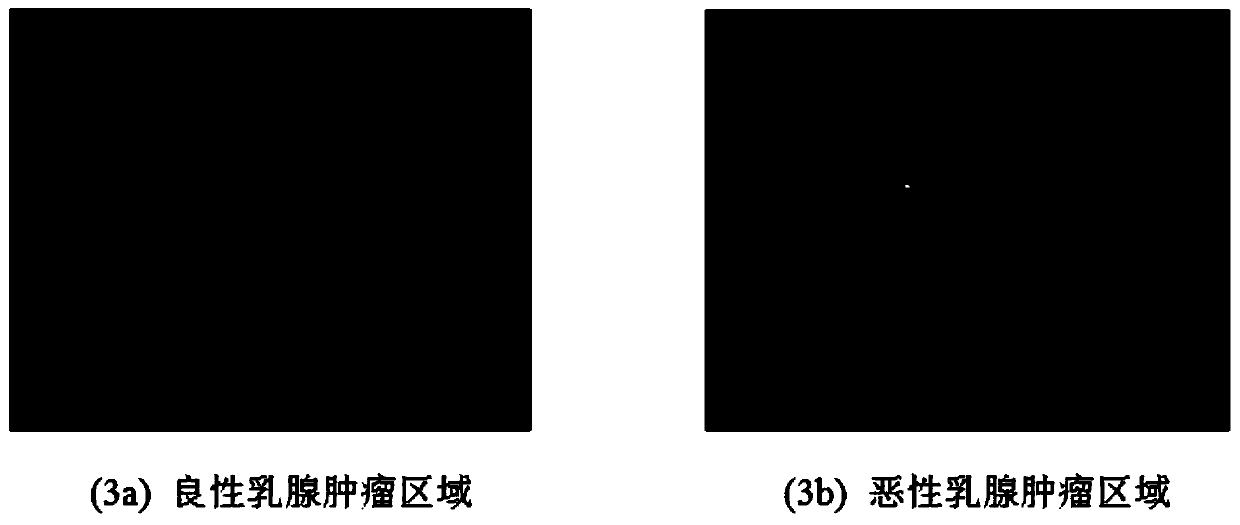
[0072] In step (A), the breast tumor region of interest is segmented from the original image of the ultrasound breast tumor image, which is segmented using the ultrasound breast tumor mask template, wherein, for example, figure 2 As shown, Figure (2a) is a benign tumor map; Figure (2b) is a malignant tumor map, such as image 3 Shown, wherein, figure (3a) is the benign breast tumor area; Figure (3b) is the malignant breast tumor area;

[0073] Step (B), distinguish between weak corrosion and strong corrosion on the segmented breast tumor region of interest, so as to reduce the influence of edge bright spots on the detection of calcification points, including the following steps...


PUM


Abstract

The invention discloses a micro calcification point automatic detection method based on an ultrasonic breast tumor image, and the method comprises the steps: segmenting an original image of the ultrasonic breast tumor image to obtain a breast tumor interest region; carrying out weak corrosion and strong corrosion distinguishing on the segmented breast tumor interest region; carrying out multi-scale superpixel segmentation on an ultrasonic breast tumor weak-corrosion interest region, and fusing the texture segmentation result of each scale to obtain a first suspected calcification point with a compact edge; performing single-scale superpixel segmentation on the original image of the ultrasonic breast tumor image, combining the original image of the ultrasonic breast tumor image after the single-scale superpixel segmentation with an ultrasonic breast tumor strong-corrosion interest region, and co-screening out a second suspected calcification point closer to the target based on the gray scale comparison characteristic and the gray scale distance characteristic; and obtaining the micro calcification point of the ultrasonic breast tumor with the accurate target and the compact edge. The method ensures that the detected tiny calcification point is accurate and reliable, is ingenious and novel to realize, and has a good application prospect.

Description

technical field [0001] The invention relates to the technical field of computer medical aided diagnosis, in particular to an automatic detection method for tiny calcification points based on ultrasound breast tumor images. Background technique [0002] At present, with the aggravation of people's fast-paced life, breast cancer is one of the malignant tumor diseases that women often suffer from, and has become the leading cause of cancer death in women. Due to changes in people's lifestyle and dietary structure, the incidence and mortality of breast cancer are increasing year by year, and the age of onset is earlier. Therefore, early detection, early diagnosis, and early treatment are very important for reducing breast cancer mortality. Ultrasound imaging has become an important means of early detection and diagnosis of breast tumors due to its advantages of no radiation trauma, low cost, real-time portability and other advantages. [0003] In BI-RADS (Breast Imaging Reporti...

Claims


Application Information

Patent Timeline
IPC IPC(8): G06T7/00G06T7/11G06T7/13G06T7/40G06T5/30G06K9/62G16H50/20
CPCG06T7/0012G06T7/13G06T7/11G06T5/30G06T7/40G16H50/20G06T2207/10132G06T2207/20104G06T2207/30068G06T2207/30096G06V10/751
Inventor 童莹任丽刘洋洋曹雪虹
Owner 南京天智信科技有限公司
Why Eureka
Try Eureka