Liver pathological image sample enhancement method based on random transformation

A technology of pathological images and samples, which is applied in image enhancement, image analysis, graphics and image conversion, etc., can solve the problems of too large sample size and insufficient sample size of pathological slices, improve reliability, and solve the problem of too large pathological slices and sample size Insufficient effect

Pending Publication Date: 2019-09-27
福州数据技术研究院有限公司
View PDF3 Cites 6 Cited by
  • Summary
  • Abstract
  • Description
  • Claims
  • Application Information

AI Technical Summary

Problems solved by technology

[0005] The purpose of the present invention is to address the deficiencies in the prior art and provide a method for enhancing liver pathology image sa

Method used


Image

  • Liver pathological image sample enhancement method based on random transformation
  • Liver pathological image sample enhancement method based on random transformation
  • Liver pathological image sample enhancement method based on random transformation

Examples


Embodiment

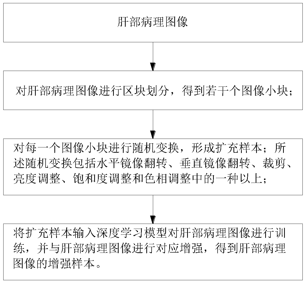
[0060] The present invention is based on random transformation liver pathological image sample enhancement method, which comprises the following steps:

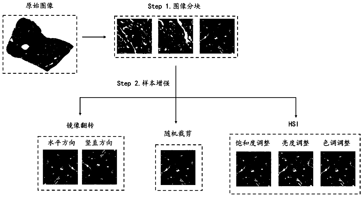
[0061]Step1, division of image blocks

[0062] Pathological images usually have extremely high resolution, and directly operating on the full image will make the calculation efficiency extremely low. Therefore, firstly, according to the resolution of the full-scan pathological image, an appropriate block length is set, and the whole pathological image is partially block. For the liver pathological image, the selected block size is 320*320 pixels, a total of wn*hn small blocks are obtained, and the position of each small block is recorded while the block is divided.

[0063] Step 2. Sample enhancement

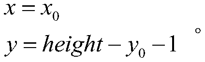
[0064] (1) Mirror flip (horizontal or vertical direction)

[0065] The principle of horizontal and vertical mirror transformation of small image blocks is as follows: Let the width of the image be width and the length be hei...


PUM


Abstract

The invention relates to a liver pathological image sample enhancement method based on random transformation, which comprises the following steps: 1) carrying out block division on a liver pathological image to obtain a plurality of image small blocks; 2) performing random transformation on each image small block to form an extended sample; wherein the random transformation comprises more than one of horizontal mirror image overturning, vertical mirror image overturning, cutting, brightness adjustment, saturation adjustment and hue adjustment; and 3) inputting the extended sample into a deep learning model to train the liver pathological image, and performing corresponding enhancement on the liver pathological image to obtain an enhanced sample of the liver pathological image. According to the method, original pathological samples can be effectively expanded, the problems of insufficient sample number and non-uniform sample distribution are solved to a certain extent, the requirement of a large sample size of a deep learning model is met, the situations of over-fitting and insufficient generalization ability of the trained model can be effectively avoided, and the reliability of an auxiliary analysis result is improved.

Description

technical field [0001] The invention relates to the technical field of digital pathological image processing, in particular to a method for segmenting liver tissue pathological images based on an OTSU threshold. Background technique [0002] Liver cancer refers to malignant tumors that occur in the liver, including primary liver cancer and metastatic liver cancer. People usually refer to primary liver cancer when they say liver cancer. Primary liver cancer is one of the most common malignant tumors in clinical practice. According to the latest statistics, there are about 600,000 new liver cancer patients in the world every year, ranking fifth among malignant tumors. There are different effective treatment methods for different cancer cell types and the extent of cancer cell spread. At present, in clinical diagnosis, especially in the diagnosis of most cancers, through biopsy, shedding and fine-needle aspiration cytology, etc., the pathological examination results obtained b...

Claims


Application Information

Patent Timeline
IPC IPC(8): G06T5/00G06T7/00G06T3/60
CPCG06T5/005G06T7/0012G06T3/60G06T2207/30056G06T2207/20021G06T2207/20081
Inventor 叶明丽林龙江雷晓晔陆长滨窦康殷陈立情
Owner 福州数据技术研究院有限公司
Why Eureka
Try Eureka