Medical image noise reduction method based on generative adversarial network and 3D residual encoding and decoding

A technology of encoding and decoding and medical images, which is applied in the field of positron emission tomography image processing, can solve the problems of easy loss of image details and slow imaging speed, etc.

Inactive Publication Date: 2019-10-01
NORTHEASTERN UNIV
View PDF4 Cites 9 Cited by
  • Summary
  • Abstract
  • Description
  • Claims
  • Application Information

AI Technical Summary

Problems solved by technology

[0004] In view of the problems in the prior art that image details are easily lost and the imaging speed is slow, the present invention provides a medical image noise reduction method based on gen

Method used


Image

  • Medical image noise reduction method based on generative adversarial network and 3D residual encoding and decoding
  • Medical image noise reduction method based on generative adversarial network and 3D residual encoding and decoding
  • Medical image noise reduction method based on generative adversarial network and 3D residual encoding and decoding

Examples


Embodiment 1

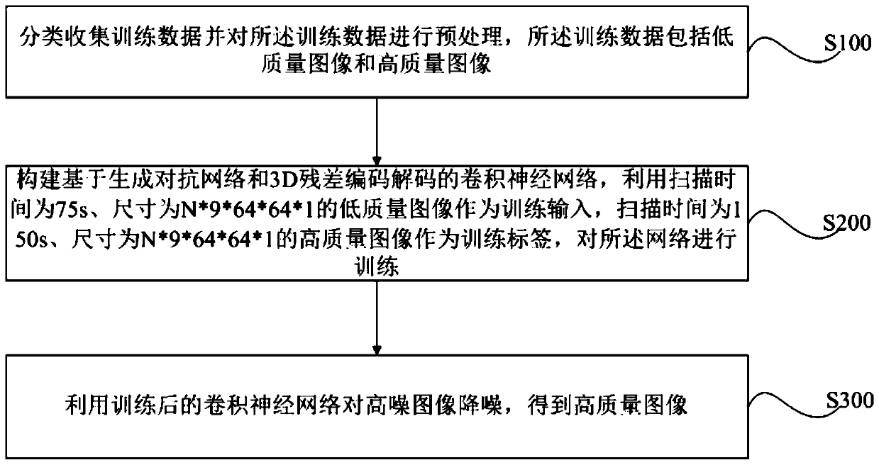
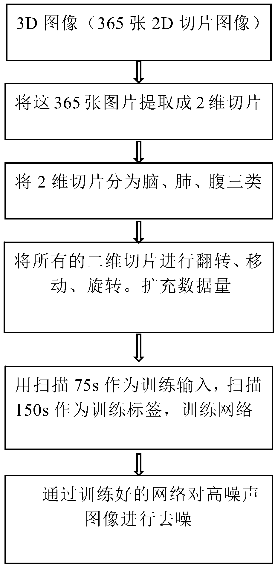
[0045] Such as figure 2 As shown, a medical image denoising method based on generative adversarial network and 3D residual coding and decoding, image denoising for positron emission tomography, including: preprocessing the collected training data; using the processed data to train Convolutional neural network based on generative confrontation network and 3D residual encoding and decoding; use the trained convolutional neural network to denoise high-noise images to obtain high-quality images.

[0046] Data preprocessing includes:

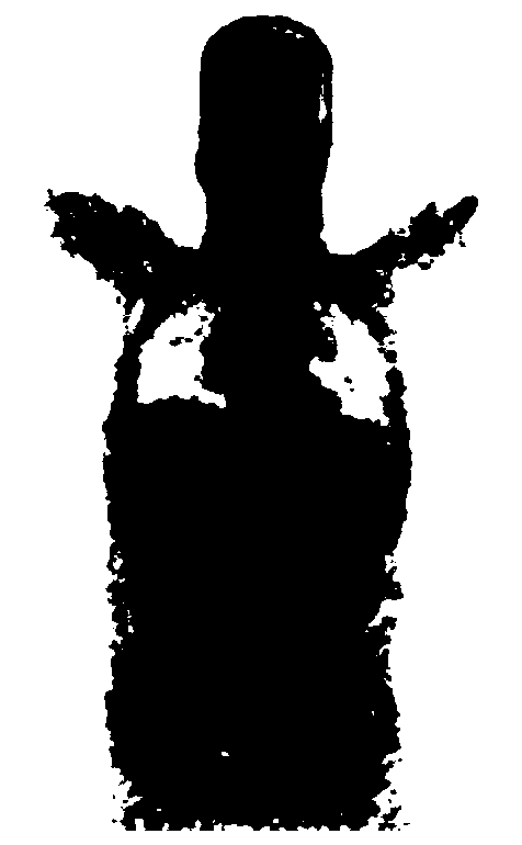
[0047] Step A: The training data is provided by Neusoft Medical, such as Figure 3a As shown, it includes a low-quality whole-body scan image with a scan time of 75s and a high-quality whole-body scan image with a scan time of 150s, and the data format is DICOM, such as Figure 3b-3d As shown, these data can be roughly divided into three categories: head, lung and abdomen.

[0048]Step B: Convert the data in DICOM format into data in npy format w...


PUM


Abstract

The invention discloses a medical image noise reduction method based on a generative adversarial network and 3D residual coding and decoding, and the method comprises the steps: carrying out the classification collection of training data, carrying out the preprocessing of the training data, wherein the training data comprises a low-quality image and a high-quality image; constructing a convolutional neural network based on a generative adversarial network and 3D residual encoding and decoding; taking a low-quality image with scanning time of 75s and size of N * 9 * 64 * 64 * 1 as training input, taking a high-quality image with scanning time of 150s and size of N * 9 * 64 * 64 * 1 as a training label, and training the network; and carrying out noise reduction on the high-noise image by using the trained convolutional neural network to obtain a high-quality image. By applying the technical scheme of the invention, any high-noise positron emission computer tomography image can be accurately and rapidly denoised after the model is trained by using a small amount of data.

Description

technical field [0001] The present invention relates to positron emission computed tomography image processing, in particular, to a medical image noise reduction method based on generative confrontation network and 3D residual coding and decoding. Background technique [0002] Positron emission tomography (PET) is a functional imaging mode that observes the molecular activity level in tissues by injecting a specific radioactive tracer. 18F-FDG is a commonly used radioactive tracer. This substance will interact with the human body Negative electrons in the tissue are annihilated, and a pair of positrons with equal energy but opposite flight directions are emitted. The detector can realize the imaging function by detecting the electron trajectory. When a disease occurs in a certain part of the human body, more active physiological activities will increase the absorption of the substance, which is different from other normal tissues. PET is widely used in clinical practice, in...

Claims


Application Information

Patent Timeline
IPC IPC(8): G06T5/00G06N3/04
CPCG06T5/002G06T2207/10104G06T2207/20081G06T2207/20084G06N3/047G06N3/045
Inventor 滕月阳龚宇
Owner NORTHEASTERN UNIV
Why Eureka
Try Eureka