
Vascular reconstruction method of MRI brain images based on 3t and 7t

A technology of blood vessel reconstruction and nuclear magnetic resonance, applied in the direction of graphic image conversion, image data processing, 2D image generation, etc., can solve the problems of scarcity, expensive 7T equipment, and difficult to reconstruct pictures.

Active Publication Date: 2021-05-14
ZHEJIANG UNIV
View PDF15 Cites 0 Cited by
  • Summary
  • Abstract
  • Description
  • Claims
  • Application Information

AI Technical Summary

Problems solved by technology

[0002] 7T magnetic resonance images can clearly see blood vessels that 3T magnetic resonance images do not have, but due to the high cost and scarcity of 7T equipment, most hospitals still use 3T images
[0007] (2) Unlike under-sampled low-resolution images and high-resolution images, 3T and 7T images are not completely registered, and it is difficult to reconstruct images directly using the differences between images

Method used


Image

  • Vascular reconstruction method of MRI brain images based on 3t and 7t
  • Vascular reconstruction method of MRI brain images based on 3t and 7t
  • Vascular reconstruction method of MRI brain images based on 3t and 7t

Examples


Embodiment 1

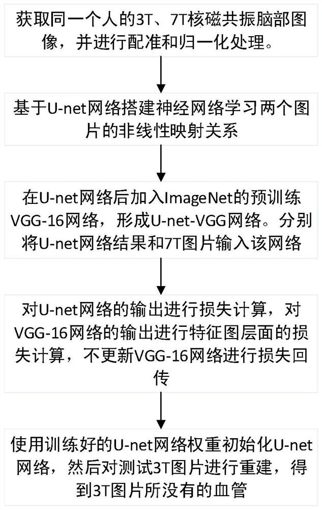
[0042] Embodiment 1, based on 3T, 7T nuclear magnetic resonance brain image blood vessel reconstruction method, such as figure 1 shown, including the following steps:

[0043] S1: Obtain multiple 3T and 7T MRI brain images of the same person, perform image preprocessing on the 3T and 7T images, and obtain normalized images;

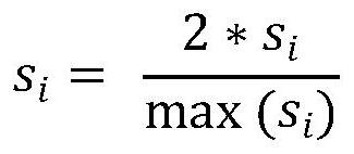
[0044] Preprocessing is to perform related operations on the image, aiming to improve its quality in order to increase the precision and accuracy of the processing algorithm in the next stage. Register the 3T and 7T MRI brain images of the same person (the original 3T and 7T images have different slice positions and angles, and the 3T and 7T images after registration are unified in angle and position, that is, each A 3T picture corresponds to a 7T picture). Afterwards for 3T pictures x 1 ,x 2 ,...x j ,...,x n and 7T picture y 1 ,y 2 ,...y j ,...,y n Each picture of is normalized, and the normalization method is shown in formula 1:

[0045]

...


PUM


Abstract

The present invention provides a 3T, 7T-based nuclear magnetic resonance brain image blood vessel reconstruction method: comprising the following steps: Step 1: Acquire 3T pictures and 7T pictures, perform image preprocessing on the 3T pictures and 7T pictures, and obtain the preprocessed 3T pictures pictures and 7T pictures; Step 2: Based on the U‑net network, use the preprocessed 3T pictures as the input of the U‑net network to obtain the output pictures through U‑net; Step 3: Separate the output pictures through U‑net Input the preprocessed 7T picture into the VGG-16 network to obtain the output of the 3T picture through the VGG-16 network and the output of the 7T picture through the VGG-16 network; Step 4: Output to the U-net network, and the 3T picture through the VGG The output after the ‑16 network and the output of the 7T image after passing through the VGG‑16 network are both calculated for loss. Based on the loss function, the parameters are obtained by using the stochastic gradient descent method, and the U‑net network is updated according to the parameters, and the 3T image is input into the updated U‑net The network gets reconstructed results. The invention can reconstruct blood vessels on 3T pictures.

Description

technical field [0001] The invention relates to the technical field of magnetic resonance imaging, in particular to a 3T and 7T-based nuclear magnetic resonance brain image blood vessel reconstruction method. Background technique [0002] 7T MRI images can clearly see blood vessels that 3T MRI images do not have, but due to the high cost and scarcity of 7T equipment, most hospitals still use 3T images. Therefore, aiming at this problem, it is very necessary to propose a vascular reconstruction method based on 3T and 7T MRI brain images to overcome this problem. [0003] Most of the existing domestic and foreign researches on image reconstruction focus on super-resolution image reconstruction. In 2014, Dong et al. proposed a CNN model SRCNN for general natural image super-resolution reconstruction. Based on SRCNN, Kim et al. proposed VDSR by referring to the VGG network structure for image classification. Bee Lim et al. proposed an enhanced deep residual network EDSR. [...

Claims


Application Information

Patent Timeline
Patent Type & Authority Patents(China)
IPC IPC(8): G06T11/00G06T3/40
CPCG06T3/4053G06T11/003
Inventor 金心宇陶建军金昀程陈智鸿
Owner ZHEJIANG UNIV
Learn More