Ankle ligament separation method and separation system in medical imaging

A ligament separation and medical imaging technology, applied in the field of medical image recognition, can solve the problem that the ankle ligament cannot be further improved in separation accuracy, and achieve the effect of improving recognition accuracy and improving recognition efficiency

Active Publication Date: 2021-02-26
北京灵医灵科技有限公司
View PDF12 Cites 0 Cited by
  • Summary
  • Abstract
  • Description
  • Claims
  • Application Information

AI Technical Summary

Problems solved by technology

[0010] In view of the above problems, the embodiment of the present invention provides a method and system for separating ankle ligaments in medical imaging, which solves the technical problem that the existing means of separating ankle ligaments cannot further improve the separation accuracy

Method used


Image

  • Ankle ligament separation method and separation system in medical imaging
  • Ankle ligament separation method and separation system in medical imaging
  • Ankle ligament separation method and separation system in medical imaging

Examples


Embodiment Construction

[0041] In order to make the purpose, technical solution and advantages of the present invention clearer and clearer, the present invention will be further described below in conjunction with the accompanying drawings and specific embodiments. Apparently, the described embodiments are only some of the embodiments of the present invention, but not all of them. Based on the embodiments of the present invention, all other embodiments obtained by persons of ordinary skill in the art without creative efforts fall within the protection scope of the present invention.

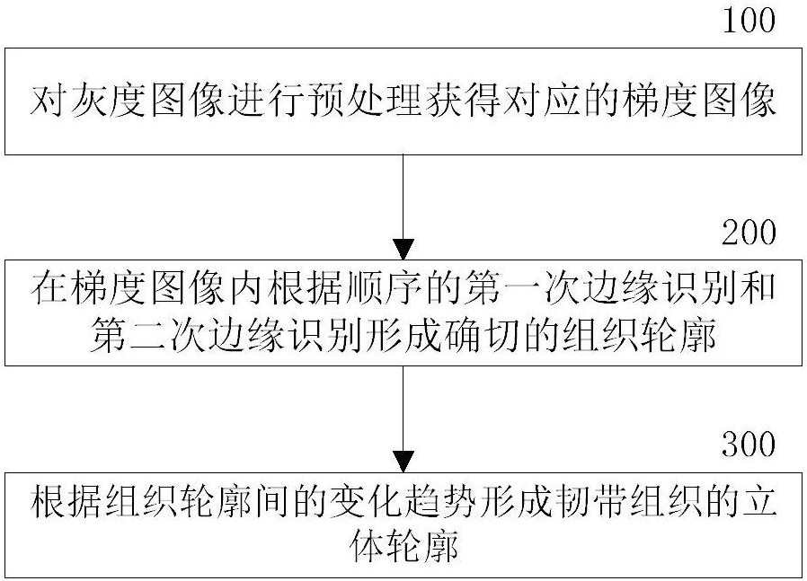
[0042] The ankle ligament separation method in an embodiment of the present invention is as follows: figure 1 shown. exist figure 1 Among them, the embodiment of the present invention includes:

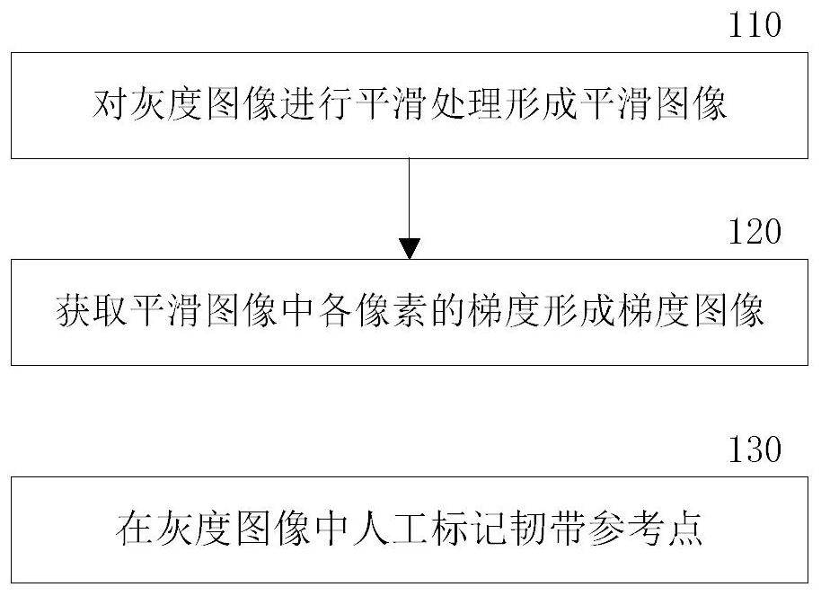
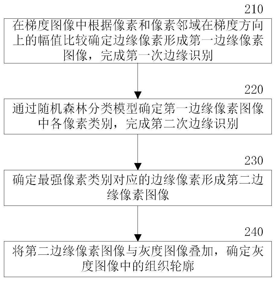
[0043] Step 100: Preprocessing the grayscale image to obtain a corresponding gradient image.

[0044] The preprocessing can obtain the quantization of a single type of information in the grayscale image, for example, the q...


PUM


Abstract

The invention provides a method and system for separating ankle ligaments in a medical image, solving the technical problem that the separation precision cannot be further improved through an existingankle ligament separation method. The method comprises the following steps: preprocessing a grayscale image to obtain a corresponding gradient image; forming an exact tissue contour in the gradient image according to sequential first edge recognition and second edge recognition; and forming a three-dimensional contour of the ligament tissue according to the change trend between the tissue contours. Automatic processing and classification are formed by utilizing mutation edges of single quantitative information, so that the tissue edge recognition accuracy is improved. Different contour generation methods are used for verification to avoid over-fitting, so that ligament tissue can realize main body automatic segmentation, contour accurate positioning and automatic three-dimensional modeling in a grayscale image, and the recognition efficiency of professional human recognition resources is effectively improved.

Description

technical field [0001] The invention relates to the technical field of medical image recognition, in particular to a method and system for separating ankle ligaments in medical images. Background technique [0002] The structure of the ankle is complex, including bones, tendons, blood vessels, and nerves. There are many small bones, and the distribution of interskeletal ligaments is dense. The contours of tissues other than bones are often unclear due to the gray-scale resolution of MRI (Magnetic Resonance Imaging, MRI), and the combination of edge gray-scale areas between tissues makes it difficult to segment ligament images [0003] In the prior art, it is necessary to manually perform selective editing, defect compensation processing, artifacts and redundant data separation on various tissue patterns, and then use a region growing method to generate segmentation results to establish a complete digital model. This will consume a lot of time for operators, and professional...

Claims


Application Information

Patent Timeline
Patent Type & Authority Patents(China)
IPC IPC(8): G06T7/12G06T5/00G06T7/13G06T7/181G06T17/00
CPCG06T5/002G06T17/00G06T2207/30008G06T7/12G06T7/13G06T7/181
Inventor 林海晓武正强
Owner 北京灵医灵科技有限公司
Why Eureka
Try Eureka