
Holistic patient radiology viewer

A technology of radiology and observers, applied in patient-specific data, promoting communication between physicians or patients, instruments, etc., can solve the time-consuming problems of medical experts

Pending Publication Date: 2019-12-03
KONINKLJIJKE PHILIPS NV
View PDF5 Cites 0 Cited by
  • Summary
  • Abstract
  • Description
  • Claims
  • Application Information

AI Technical Summary

Problems solved by technology

However, this is time consuming for medical specialists, and furthermore not all medical specialists are proficient at interpreting complex medical findings in a way that can be easily understood by lay patients

Method used


Image

  • Holistic patient radiology viewer
  • Holistic patient radiology viewer
  • Holistic patient radiology viewer

Examples


Embodiment Construction

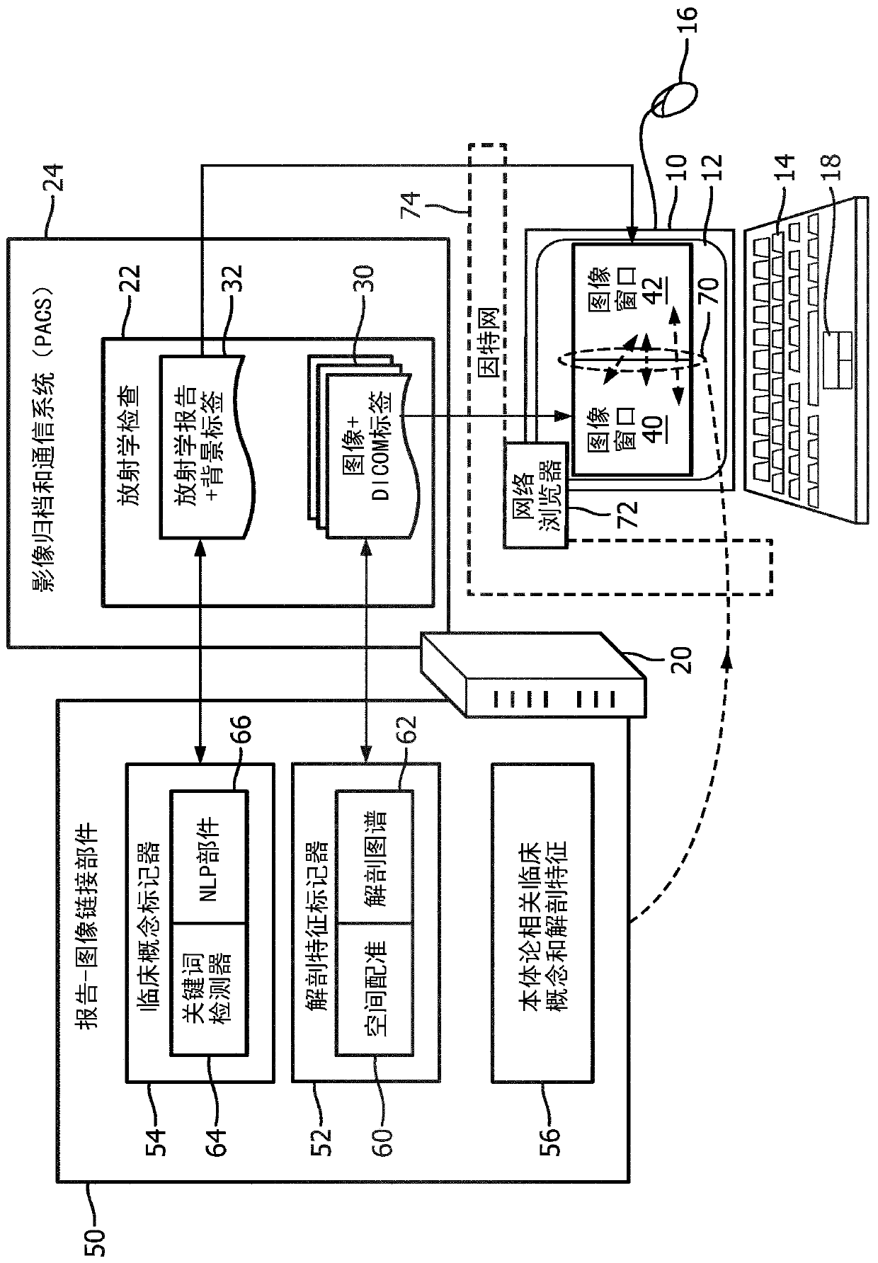
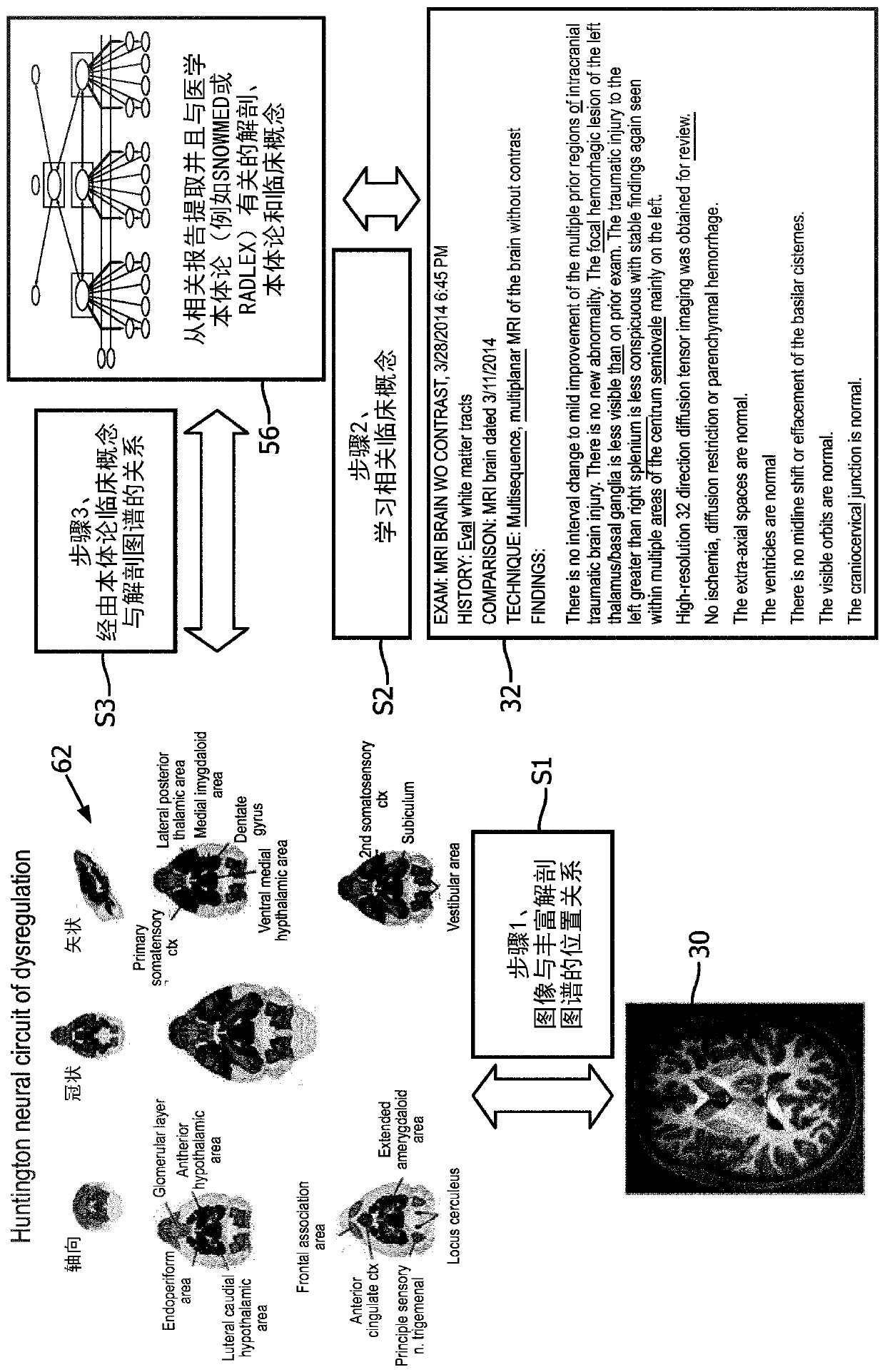
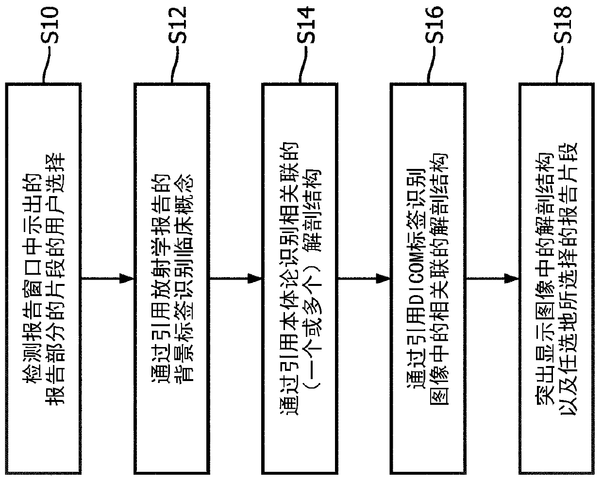
[0018] Disclosed herein is a radiology method for determining the links between clinical concepts present in a radiology report of a radiology examination and the relevant anatomical features in an underlying medical image and graphically presenting these links to the patient or other user in an intuitive manner. Learning Observer. The disclosed improvement is proposed in part on the realization that interpretation of the results of radiological examinations often requires synthesis of the content of radiological reports with features shown in the underlying medical images.

[0019] In the case where the user is a lay patient or otherwise, it should further be recognized that the user may often not be familiar with the anatomical context of the clinical findings reported in radiology - thus the disclosed radiology viewer presents selected features by selecting the context Interpreting and highlighting any associated content of the radiology report provides for the user to iden...


PUM


Abstract

A radiology viewer includes at least one electronic processor (10, 20), at least one display (12), and at least one user input device (14, 16, 18). The display shows at least a portion of a radiologyimage (30) in an image window (40), and at least a portion of a radiology report (32) in a report window (42). A selection is received of an anatomical feature shown in the image window, and a corresponding passage of the radiology report is identified and highlighted in the report window. A selection is received of a passage of the radiology report shown in the report window, and a correspondinganatomical feature of the at least one radiology image is identified and highlighted in the image window. The highlighting operations use image anatomical feature tags and report clinical concept tagsgenerated using a medical ontology (56) and an anatomical atlas (62).

Description

technical field [0001] The following generally relates to the field of medical imaging, medical image viewers and displays, and related fields. Background technique [0002] In modern medical practice, the patient is expected to be an active participant in his or her medical care. For example, patients must provide informed consent to various medical processes if they are physically and mentally able to do so. To this end, it is important that the patient understands the findings of a medical examination, such as a radiological examination. [0003] However, most lay patients (ie, patients without medical training) are not familiar with detailed anatomy, let alone visualization of such anatomy as present in medical images. In a typical radiology workflow, images of a radiology exam are interpreted by a skilled radiologist who prepares a radiology report outlining the radiologist's clinical findings. However, radiology reports use high-level clinical language and anatomica...

Claims


Application Information

Patent Timeline
IPC IPC(8): G16H15/00G16H30/40G16H80/00
CPCG16H30/40G16H80/00G16H15/00G16H10/60G16H30/20G16H50/70
Inventor N·沙德瓦尔特A·M·塔赫玛塞比马拉古奥施L·G·扎戈尔谢夫S·M·达拉尔D·贝斯特罗夫A·R·弗兰茨
Owner KONINKLJIJKE PHILIPS NV
Learn More