DR image pulmonary tuberculosis intelligent segmentation and detection method based on deep learning

A deep learning and detection method technology, applied in the medical field, can solve the problems of high recognition rate, DR chest X-ray output, etc., to achieve the effect of accurate output, efficient reference, and improved detection rate

Inactive Publication Date: 2020-02-11
ZHEJIANG UNIV
View PDF6 Cites 3 Cited by
  • Summary
  • Abstract
  • Description
  • Claims
  • Application Information

AI Technical Summary

Benefits of technology

This technology helps identify people who have been exposed to active disease by analyzing X-ray images that show suspicious lungs regions within their body's organs. By comparing this analysis data against previous results collected during routine medical examinations, we aimed at identifying patients likely to be sick because they were infected while being treated earlier than usual. It could help prevent unnecessary treatments like antibiotic therapy without actually showing any symptoms.

Problems solved by technology

This patented technical solution describes how identifies suspected individuals from CT scans can be useful tools used during clinic visits due to their low incidence compared to other methods such as x ray film analysis.

Method used


Image

  • DR image pulmonary tuberculosis intelligent segmentation and detection method based on deep learning
  • DR image pulmonary tuberculosis intelligent segmentation and detection method based on deep learning

Examples


Embodiment 1

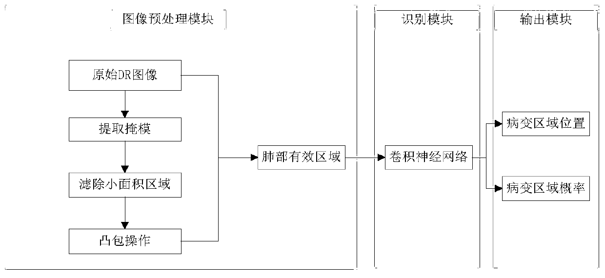
[0025] A method for intelligent segmentation and detection of pulmonary tuberculosis in DR images based on deep learning, such as figure 1 shown, including the following steps:

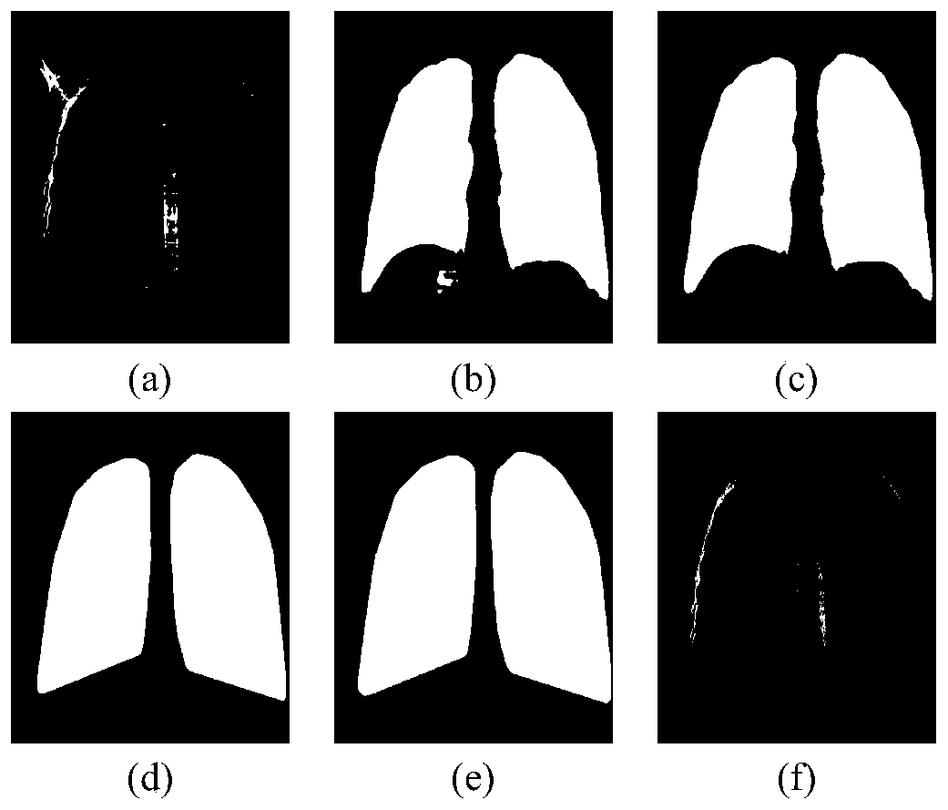
[0026] (1) Normalize the original DR image pixel value to 0-255, see figure 2 middle (a);

[0027] (2) Use the convolutional neural network to segment the above image, extract the mask of the effective area of ​​the lung, and obtain the binary image of the effective area of ​​the lung, see figure 2 middle (b);

[0028] (3) Filter out the small area of ​​the mask image extracted in the previous step, see figure 2 middle (c);

[0029] (4) Perform convex hull operation on the mask in the previous step, see figure 2 middle (d);

[0030] (5) Perform an expansion operation on the mask after the convex hull operation to expand the effective area of ​​​​the lungs, see figure 2 middle (e);

[0031] (6) Multiply the images obtained in step (1) and step (5) to obtain the effective area of ​​the lung...


PUM


Abstract

The invention discloses a DR image pulmonary tuberculosis intelligent segmentation and detection method based on deep learning, and the method comprises the steps: extracting a mask from an original DR image, filtering a small-area region, carrying out convex hull operation, and carrying out texpansion operation of the mask after the convex hull operation, and obtaining a lung effective region; labeling the original DR image, and labeling the position of a lesion area of the DR image with pulmonary tuberculosis expression; sending the masked image and the annotation information containing thepulmonary tuberculosis change area position into a deep neural network for training, and learning features of the pulmonary tuberculosis image; and using the trained model to predict the untrained DRimage, outputting the position of the pulmonary tuberculosis change area, and giving the probability of the pulmonary tuberculosis change area. According to the invention, the detection rate of pulmonary tuberculosis is greatly improved, the position of a pulmonary tuberculosis change area is accurately output, and reliable and efficient reference is provided for screening and treatment of pulmonary tuberculosis.

Description


Claims


Application Information

Patent Timeline
Owner ZHEJIANG UNIV
Why Eureka
Try Eureka