Methods of isolating T cells having antigenic specificity for P53 cancer-specific mutation

A specific and antigen-specific technology, applied in the field of isolating T cells with antigen specificity for P53 cancer-specific mutations, can solve the problems of difficult identification and isolation of T cells

Pending Publication Date: 2020-06-26
UNITED STATES OF AMERICA
View PDF9 Cites 2 Cited by
  • Summary
  • Abstract
  • Description
  • Claims
  • Application Information

AI Technical Summary

Problems solved by technology

For example, T cells that specifically recognize cancer antigens can be difficult to identify and / or isolate from patients

Method used


Image

  • Methods of isolating T cells having antigenic specificity for P53 cancer-specific mutation
  • Methods of isolating T cells having antigenic specificity for P53 cancer-specific mutation
  • Methods of isolating T cells having antigenic specificity for P53 cancer-specific mutation

Examples


Embodiment 1

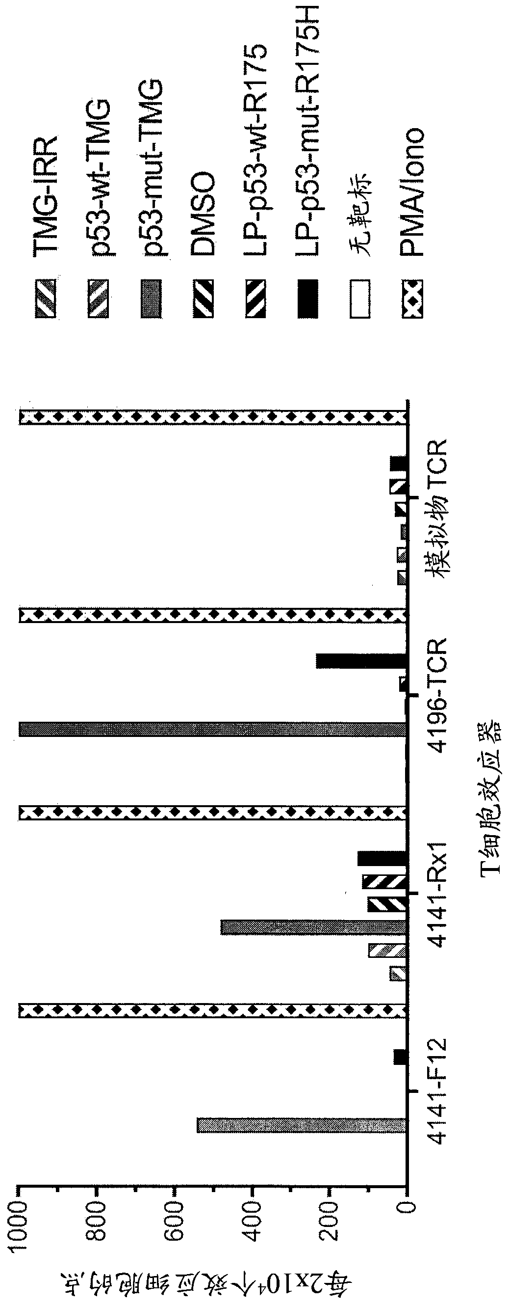
[0139] This example demonstrates the identification of anti-mutant p53 T cells in patient 4141 by co-culturing autologous APCs induced to express mutated p53 within autologous T cells ("p53 hotspot mutation universal screen"). This example also demonstrates the isolation and specific reactivity of the TCR from patient 4141.

[0140] Such as figure 1 and 45 -48 Experiments were performed as described for patient 4141. TIL fragment F12 and infusion bag TIL (Rx1 ) from patient 4141 and p53-R175H-specific TCR or mock-transduced T cells from patient 4196 were used as effectors. Co-cultures of T cell effectors with HLA-A*02:01APCs (autologous for patient 4141) were (1) electroporated with TMG consisting of irrelevant, WT p53, or mutated p53 sequences, or (2) with a peptide vehicle (DMSO) or a purified (>95% by HPLC) 25 amino acid peptide pulse consisting of the WT p53-R175 sequence or the mutated p53-R175H sequence. T cells only (no target) are negative controls, and PMA and Ion...

Embodiment 2

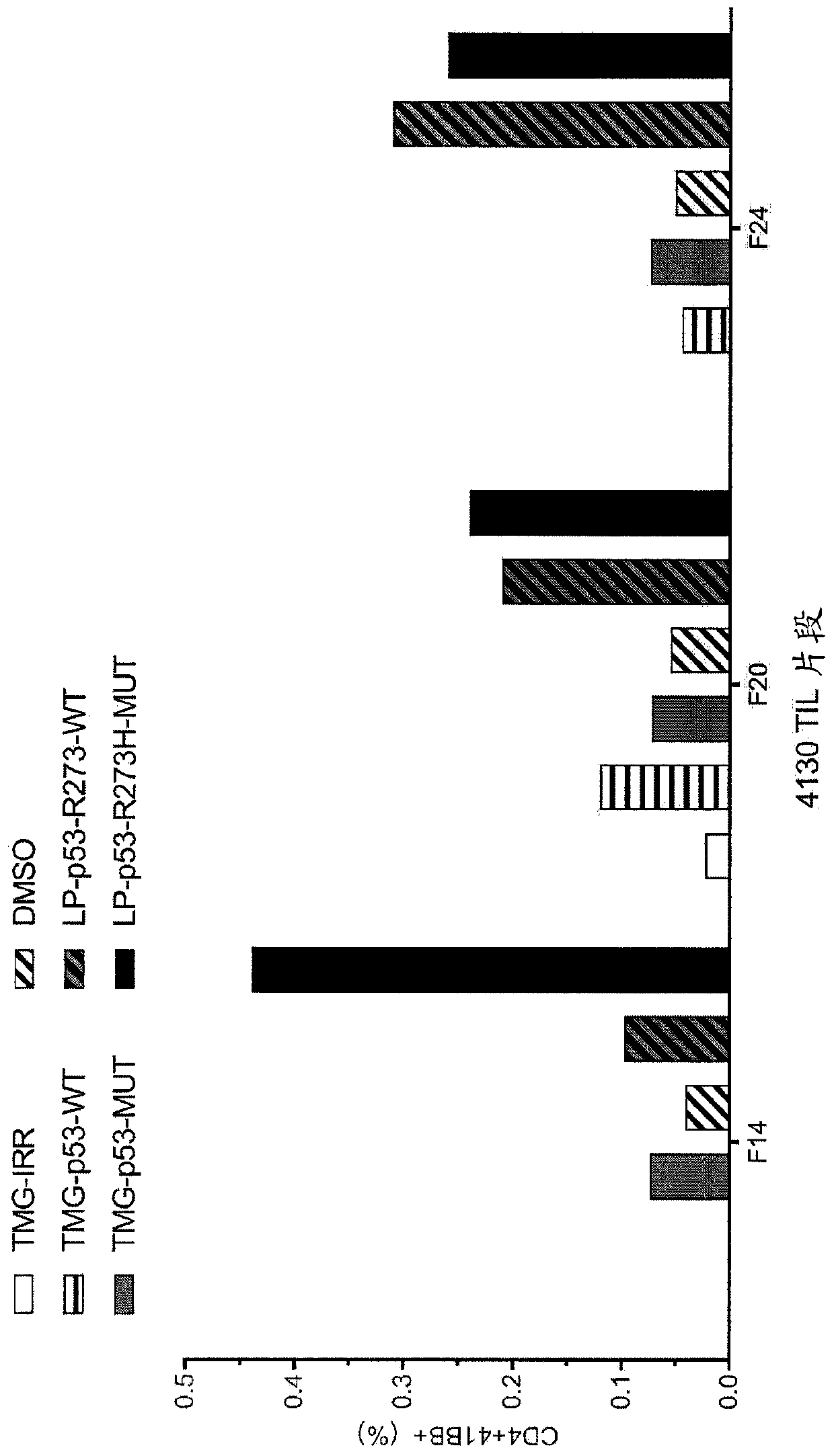
[0159] This example demonstrates the identification of anti-mutant p53 T cells in patient 4130 by co-culturing autologous APCs induced to express mutated p53 within autologous T cells ("p53 hotspot mutation universal screen").

[0160] Such as figure 2 Experiments were performed as described for patient 4130. TIL fragments (F14, F20 and F24) from patient 4130 were co-cultured with autologous APCs (1) electroporated with TMG consisting of irrelevant, WT p53 or mutated p53 sequences, or (2) Pulse with peptide vehicle (DMSO) or purified (>95% by HPLC) 25 amino acid peptide consisting of WT p53-R273 sequence or mutated p53-R273H sequence. Co-cultivation was performed overnight at 37°C. Expression of 4-1BB was assessed by flow cytometry after gating on lymphocytes→viable cells (PI negative)→CD3+ (T cells). The results are shown in figure 2 middle.

Embodiment 3

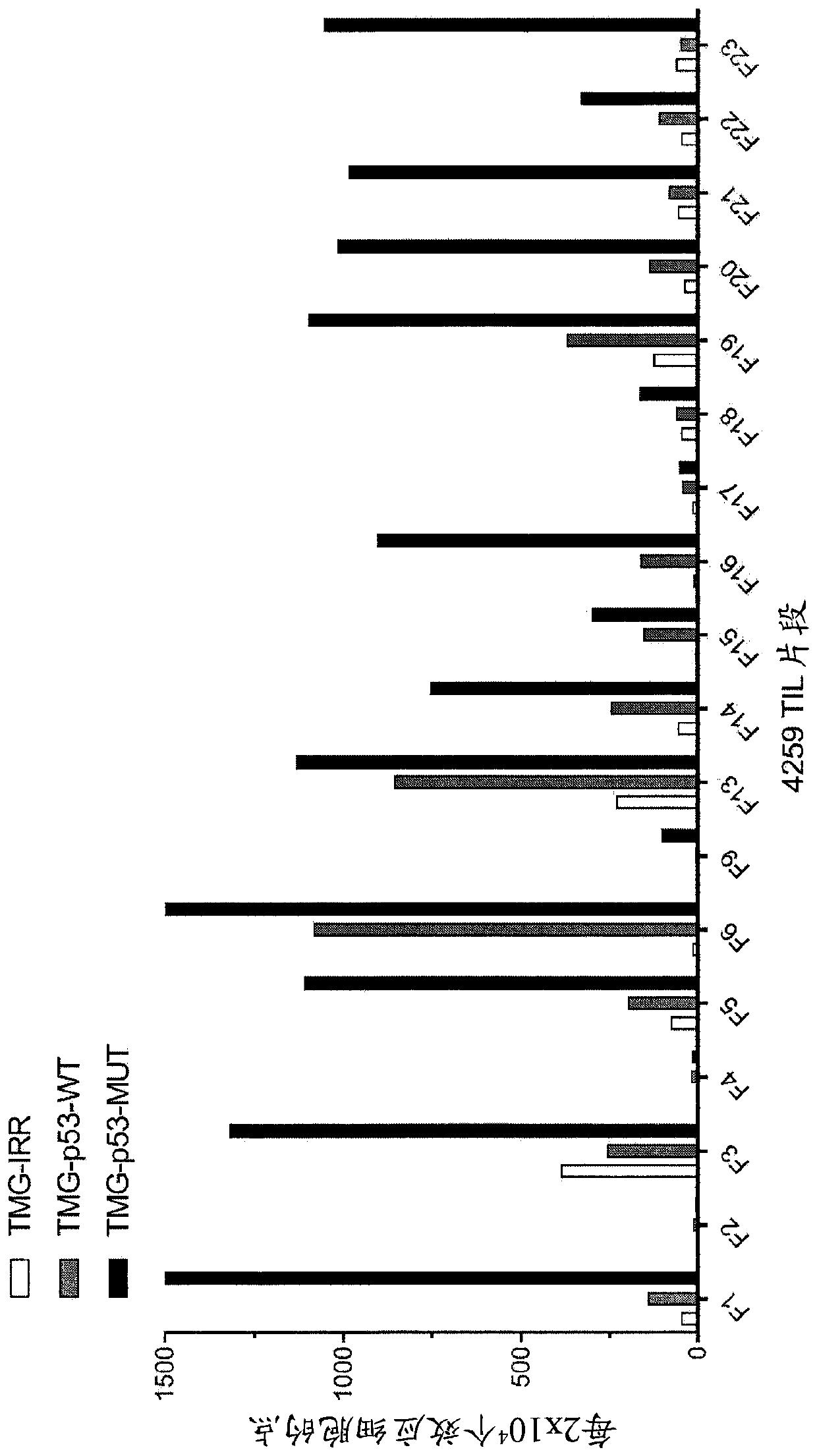
[0162] This example demonstrates the identification of anti-mutant p53 T cells in patient 4259 by co-culturing autologous APCs induced to express mutated p53 within autologous T cells ("p53 hotspot mutation universal screen"). This example also demonstrates the isolation and specific reactivity of the TCR isolated from patient 4259.

[0163] Such as Figure 3-8 and 49-53 were performed as described for patient 4259. TIL fragments (n=18) from patient 4259 were co-cultured with autologous APCs electroporated with TMG consisting of irrelevant, WT p53 or mutated p53 sequences. Co-cultivation was performed overnight at 37°C. IFN-γ secretion was assessed using an ELISPOT assay. The results are shown in image 3 middle. Expression of 4-1BB was assessed by flow cytometry after gating on lymphocytes→viable cells (PI negative)→CD3+ (T cells). The results are shown in Figure 4-5 middle.

[0164] TIL fragments (n=18) from patient 4259 were combined with a purified (>95% by HPLC) 25...


PUM


Abstract

Disclosed are methods of isolating T cells having antigenic specificity for a mutated p53 amino acid sequence encoded by a cancer-specific p53 mutation, the method comprising: inducing autologous APCsof the patient to present the mutated p53 amino acid sequence; co-culturing autologous T cells of the patient with the autologous APCs that present the mutated p53 amino acid sequence; and selectingthe autologous T cells. Also disclosed are related methods of preparing a population of cells, populations of cells, pharmaceutical compositions, and methods of treating or preventing cancer.

Description

[0001] Cross References to Related Applications [0002] This patent application claims the benefit of U.S. Provisional Patent Application No. 62 / 565,464, filed September 29, 2017, which is hereby incorporated by reference in its entirety. [0003] Statement Regarding Federally Funded Research and Development [0004] This invention was made with government support from National Institutes of Health, National Cancer Institute (National Institutes of Health, National Cancer Institute), Project No. ZIABC010984. The government has certain rights in this invention. [0005] Incorporation by reference of electronically submitted material [0006] Incorporated herein in its entirety by reference is the computer readable nucleotide / amino acid sequence listing filed at the same time and identified as follows: A 687,616 byte ASCII ( text) file. [0007] Background of the invention [0008] Adoptive cell therapy (ACT) can produce positive clinical responses in certain cancer patients...

Claims


Application Information

Patent Timeline
Patent Type & Authority Applications(China)
IPC IPC(8): C12N5/0783C12N5/0784A61K35/17A61K39/00C07K14/47
CPCA61K35/17C07K14/4746C12N5/0636C12N2502/1121A61K39/001151A61P35/00C07K14/47
Inventor 德鲁·C·丹尼格尔史蒂文·A·罗森伯格帕里萨·马勒克扎德
Owner UNITED STATES OF AMERICA
Why Eureka
Try Eureka