
Automatic skull removal method for brain magnetic resonance image

A magnetic resonance image and brain technology, applied in image enhancement, image analysis, image data processing, etc., can solve the problem of difficulty in selecting the global threshold of brain magnetic resonance images, and achieve automatic identification and image fusion. Time complexity, strong applicability

Active Publication Date: 2020-09-01
SHENYANG INST OF AUTOMATION - CHINESE ACAD OF SCI
View PDF2 Cites 4 Cited by
  • Summary
  • Abstract
  • Description
  • Claims
  • Application Information

AI Technical Summary

Problems solved by technology

[0003] Aiming at the deficiencies of the prior art, the present invention provides an automatic skull removal method for brain magnetic resonance images, which solves the problem that it is difficult to select a global threshold for all brain magnetic resonance images due to non-standardization of brain magnetic resonance images

Method used


Image

  • Automatic skull removal method for brain magnetic resonance image
  • Automatic skull removal method for brain magnetic resonance image
  • Automatic skull removal method for brain magnetic resonance image

Examples


Embodiment

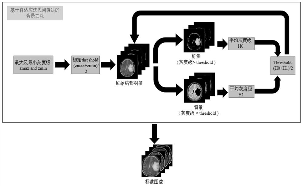
[0053] Taking the brain magnetic resonance image of the T2 weighted sequence as an example, after obtaining the brain magnetic resonance image set, in order to facilitate subsequent operations, it is necessary to perform background removal on the brain magnetic resonance image first. Due to the large gap between the background and foreground gray levels, the present invention uses an adaptive iterative threshold method to achieve background removal. The principle of the adaptive iterative threshold method is as follows: figure 2 As shown, the average of the highest and lowest gray levels in the magnetic resonance image is selected as the initial threshold, and the image is divided into foreground and background according to the initial threshold; then the average gray level of the foreground and background is calculated, and the average gray level of the foreground and background The average number of levels is used as the new threshold, and the original image is divided into ...


PUM


Abstract

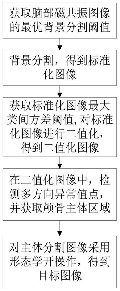
The invention relates to an automatic skull removal method for a brain magnetic resonance image. The method comprises: obtaining an optimal background segmentation threshold of the brain magnetic resonance image through an adaptive iterative threshold method; performing background segmentation on the brain magnetic resonance image according to the optimal background segmentation threshold to obtain a standardized image; obtaining a maximum inter-class variance threshold of the standardized image through an OSTU threshold method, and binarizing the standardized image according to the maximum inter-class variance threshold to obtain a binarized image; in the binarized image, detecting multi-directional abnormal value points, and obtaining a skull main body area; in the standardized image, assigning 0 to the pixels of the skull main body area to obtain a main body segmentation image; and performing morphological opening operation on the main body segmentation image to obtain a target image. According to the invention, the efficiency of a traditional semi-automatic method is effectively improved, and convenience is provided for operations such as automatic identification and image fusion of brain magnetic resonance images.

Description

technical field [0001] The invention relates to the field of medical image segmentation, in particular to an automatic skull removal method for brain magnetic resonance images. Background technique [0002] Since the skull has a gray distribution similar to that of brain tumors and other brain tissues in the brain magnetic resonance image, the presence of the skull will greatly interfere with the processing of brain magnetic resonance images such as automatic diagnosis of brain tumors and image fusion. Skull removal is required on MRI images of the brain. Traditional skull removal methods mostly use the global threshold method and region growing method. The global threshold method has poor applicability. Due to the non-standardization of brain magnetic resonance images, it is difficult to select a global threshold for all brain magnetic resonance images. In addition, the region growing method requires manual Selecting seed points is a semi-automatic segmentation method with...

Claims


Application Information

Patent Timeline
IPC IPC(8): G06T7/11G06T7/136G06T7/155G06T7/194
CPCG06T7/194G06T7/136G06T7/155G06T7/11G06T2207/10088G06T2207/30008Y02A90/30
Inventor 宋国立黄钲赵忆文赵新刚
Owner SHENYANG INST OF AUTOMATION - CHINESE ACAD OF SCI
Learn More