Method and apparatus to detect lesions of diabetic retinopathy in fundus images

a technology of diabetic retinopathy and fundus, which is applied in the field of method and apparatus to detect lesions of diabetic retinopathy in fundus images, can solve the problems of insufficient referral, economic hindrance, and insufficient access to proper eye care, and achieves the effects of reducing time complexity, reducing run-time complexity, and fast dr detection system

Inactive Publication Date: 2014-10-23
PARHI KESHAB K +1
View PDF6 Cites 43 Cited by
  • Summary
  • Abstract
  • Description
  • Claims
  • Application Information

AI Technical Summary

Benefits of technology

[0007]One aspect of the proposed invention is the 3-stage system design where each stage has minimal run-time complexity to ensure a fast DR detection system. An optimal feature set is defined that will allow classifiers to detect retinopathy lesions and to generate a severity grade for a fundus image (See, S. Roychowdhury, D. Koozekanani, and K. K. Parhi, “DREAM: Diabetic Retinopathy Analysis using Machine Learning,” IEEE Journal of Biomedical and Health Informatics, 2014, doi: 10.1109JBHI.2013.2294635).
[0008]A key contribution of the proposed invention is a novel two-step hierarchical binary classification method that rejects false positives in the first step and in the second step, bright lesions are classified as cotton wool spots (CWS) or hard exudates (HE), and red lesions are classified as hemorrhages (HA) and micro-aneurysms (MA), respectively. This hierarchical classification method reduces the time complexity by 18-24% over a parallel classification method that trains separate classifiers for identifying CWS, HE, HA and MA from false positives.

Problems solved by technology

Unfortunately almost 50% of diabetic patients in the United States currently do not undergo any form of documented screening exams in spite of the guidelines established by the American Diabetes Association (ADA) and the American Academy of Ophthalmology (AAO).
The major reasons for this screening and treatment gap include insufficient referrals, economic hindrances and insufficient access to proper eye care.

Method used


Image

  • Method and apparatus to detect lesions of diabetic retinopathy in fundus images
  • Method and apparatus to detect lesions of diabetic retinopathy in fundus images
  • Method and apparatus to detect lesions of diabetic retinopathy in fundus images

Examples


examples

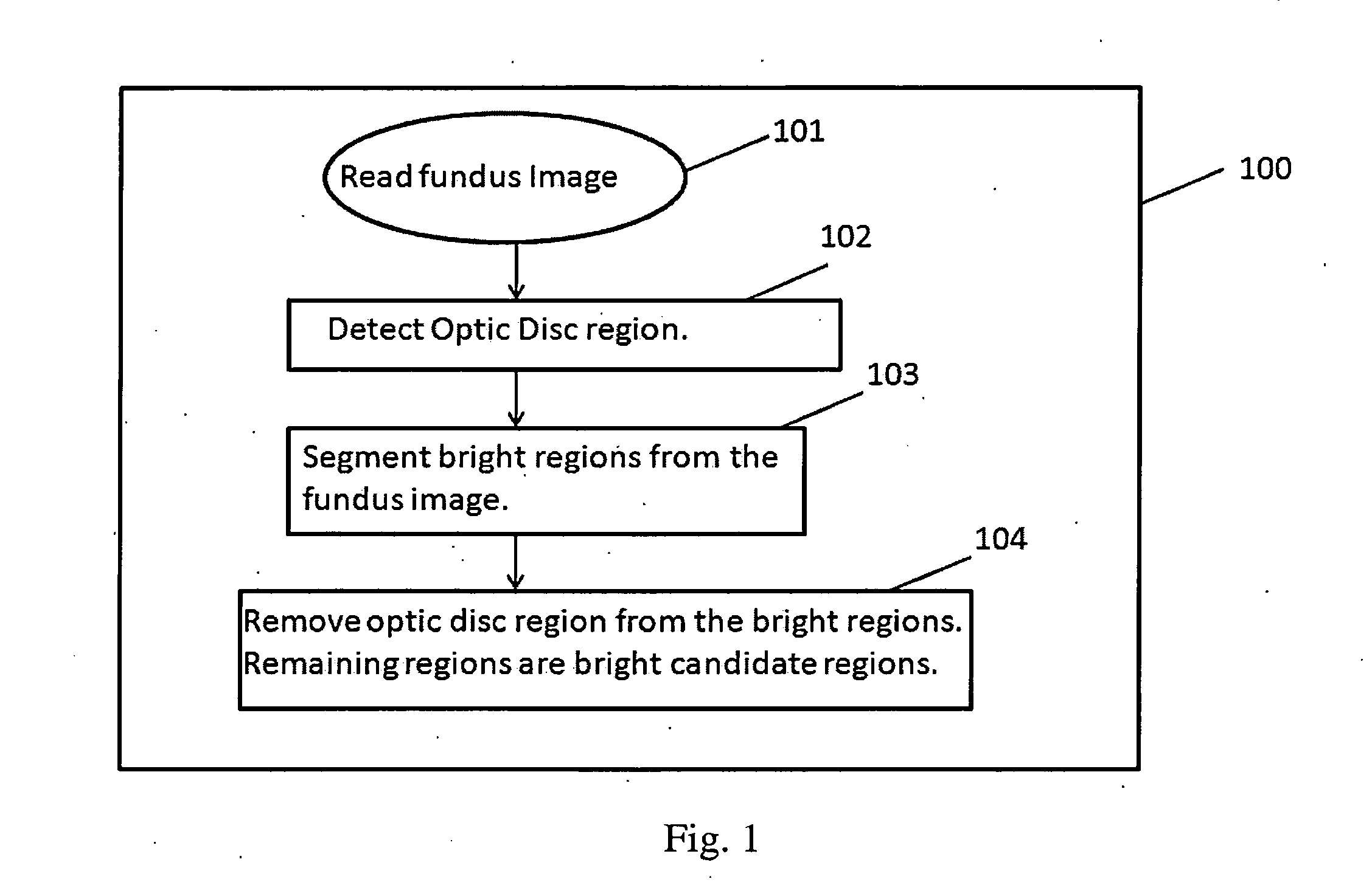
[0043]The three stages of the proposed invention are illustrated with an example in FIG. 7, FIG. 8, FIG. 9 and FIG. 10. The first stage involving extraction of bright and red candidate regions is shown in FIG. 7 and FIG. 8, respectively. FIG. 7A shows the fundus image received. FIG. 7B shows the outcome of the automated OD region detection algorithm superimposed on the original image. Next, the OD region is removed from the bright regions detected from the image, and the remaining bright candidate regions (RBR) superimposed on the green plane of the fundus image are shown in FIG. 7C. The pixels marked in white in FIG. 7C represent the bright candidate regions.

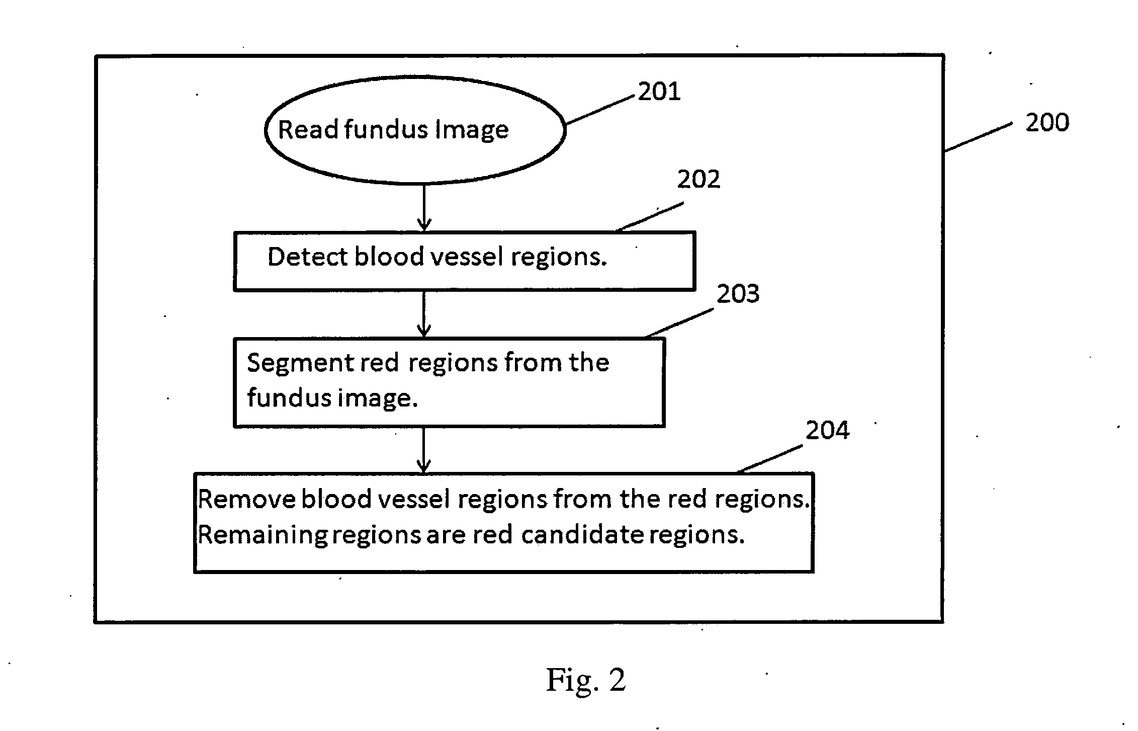
[0044]FIG. 8A shows the same fundus image as in FIG. 7A. FIG. 8B shows the blood vessel regions detected and FIG. 8C shows the red candidate regions (RRR) after removing the blood vessel regions from the red regions.

[0045]The second stage of the proposed invention involving classification of the bright and red candidate regions...


PUM


Abstract

The present invention relates to the design and implementation of a three stage computer-aided screening system that analyzes fundus images with varying illumination and fields of view, and generates a severity grade for diabetic retinopathy (DR) using machine learning. In the first stage, bright and red regions are extracted from the fundus image. An optic disc has similar structural appearance as bright lesions, and the blood vessel regions have similar pixel intensity properties as the red lesions. Hence, the region corresponding to the optic disc is removed from the bright regions and the regions corresponding to the blood vessels are removed from the red regions. This leads to an image containing bright candidate regions and another image containing red candidate regions. In the second stage, the bright and red candidate regions are subjected to two-step hierarchical classification. In the first step, bright and red lesion regions are separated from non-lesion regions. In the second step, the classified bright lesion regions are further classified as hard exudates or cotton-wool spots, while the classified red lesion regions are further classified as hemorrhages and micro-aneurysms. In the third stage, the numbers of bright and red lesions per image are combined to generate a DR severity grade. Such a system will help in reducing the number of patients requiring manual assessment, and will be critical in prioritizing eye-care delivery measures for patients with highest DR severity.

Description

CROSS-REFERENCE TO RELATED APPLICATIONS[0001]This application claims the benefit of U.S. Provisional Application No. 61 / 854,034, filed on Apr. 17, 2013, the entire content of which is incorporated herein by reference in its entirety.FIELD OF THE INVENTION[0002]Automated detection of diabetic retinopathy (DR) lesions from fundus images is important for detecting ophthalmic abnormalities and for developing cost-effective DR screening systems that will help in grading severity of non-proliferative DR. This will enhance the effectiveness of the present day eye-care delivery.BACKGROUND OF THE INVENTION[0003]According to a study by the American Diabetes Association, diabetic retinopathy (DR) had affected more than 4.4 million Americans of age 40 and older during 2005-2008, with almost 0.7 million (4.4% of those with diabetes) having advanced DR that could lead to severe vision loss. Early detection and treatment of DR can provably decrease the risk of severe vision loss by over 90%. Thus,...

Claims


Application Information

Patent Timeline
Patent Type & Authority Applications(United States)
IPC IPC(8): G06T7/00A61B3/12
CPCA61B3/1241G06T7/0012G06T2207/30041G06T7/11
Inventor ROYCHOWDHURY, SOHINIPARHI, KESHAB K.KOOZEKANANI, DARA D.
Owner PARHI KESHAB K
Why Eureka
Try Eureka