
Method for producing a tailor-made implant

A manufacturing method and implant technology, applied in the direction of joint implants, joint implants, general control systems, etc., can solve the problems of insufficient transmission force, unsatisfactory, and not allowing practitioners joint freedom, etc. To achieve the effect of ensuring healthy distribution, saving bone mass and saving time

Inactive Publication Date: 2020-10-30
3D MEDICAL
View PDF9 Cites 2 Cited by
  • Summary
  • Abstract
  • Description
  • Claims
  • Application Information

AI Technical Summary

Problems solved by technology

[0013] Furthermore, in the case of extensive and substantially planar resections, the support of the prosthesis on the resected surface is unsatisfactory because this surface is essentially composed of cancellous bone, and the support on the cortical periphery of the bone plays an important role in transmitting forces. Insufficient aspects
[0014] Finally, known implants do not allow the practitioner to choose from a sufficient range to satisfy the selected joint degrees of freedom, said implants being standardized

Method used


Image

  • Method for producing a tailor-made implant
  • Method for producing a tailor-made implant
  • Method for producing a tailor-made implant

Examples


Embodiment Construction

[0171] First, it should be noted that the drawings are not drawn to scale.

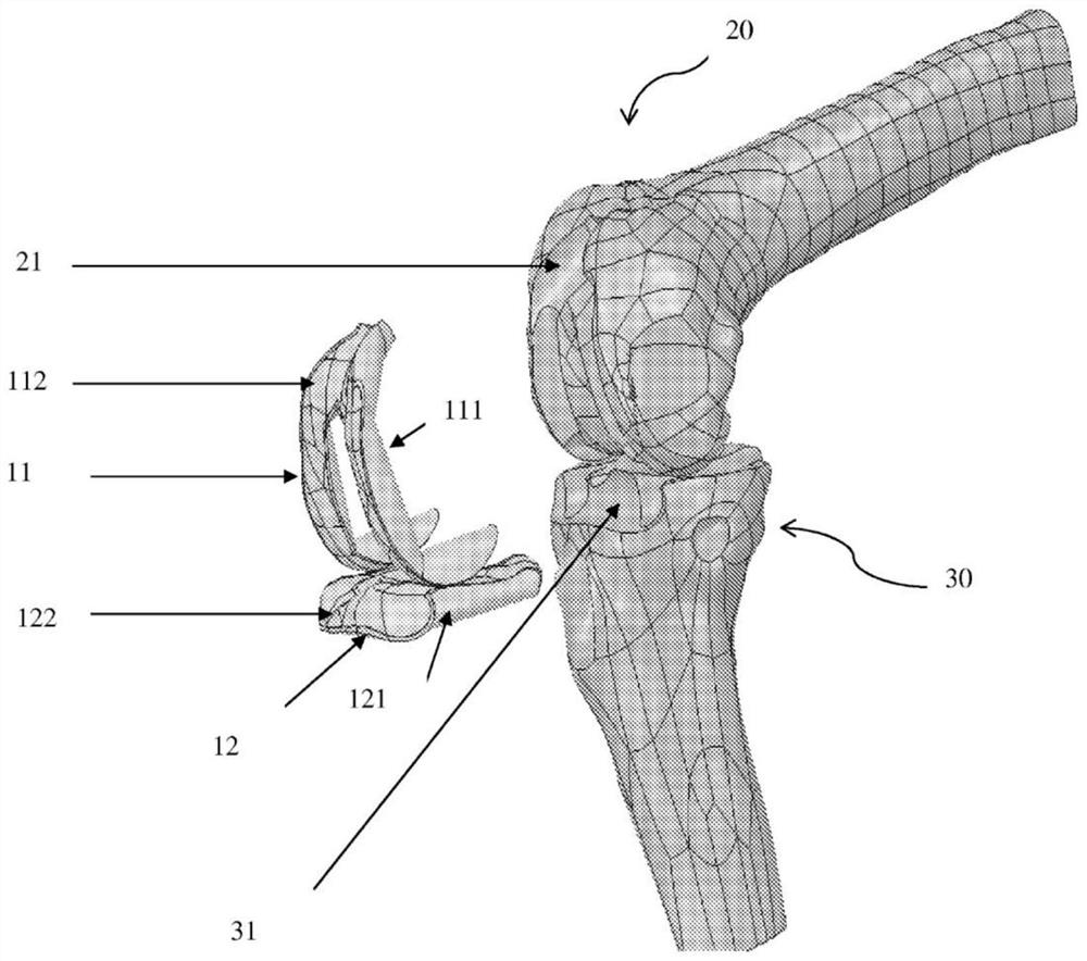
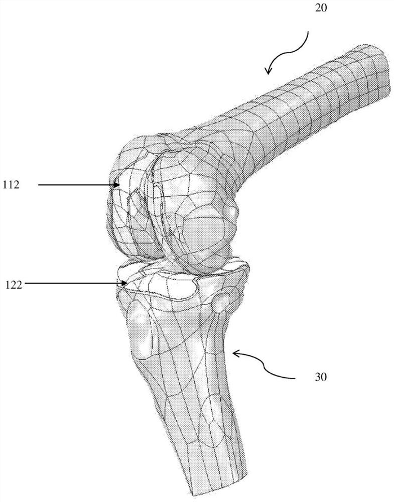
[0172] An image of the skeletal part (here the skeletal part fitting in the tibiofemoral joint) has been acquired by magnetic resonance imaging, and the image has then been segmented and 3D modeled on a computer, which is transferred in STL format. Thus, a 3D image of the tibiofemoral joint (i.e. of the proximal end 30 of the tibia and the distal end 20 of the femur) has been acquired, and e.g. figure 1 As shown, the image is located in a functional position.

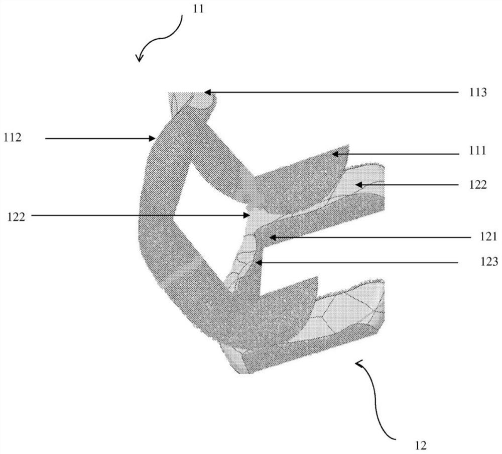
[0173] On the basis of the 3D image of the tibiofemoral joint in functional position, the recesses required to accommodate the custom implants 11, 12 have been made on the 3D image of the tibiofemoral joint in order to locally modify the proximal tibial end 30 and Distal femoral end 20 .

[0174] A recess 21 has been formed on the distal femoral end 20 and a recess 31 has been formed on the proximal tibial end 30 .

[0175] By means of each ...


PUM


Abstract

The present invention relates to a method for producing a tailor-made implant intended to be implanted at an implantation site of a damaged bone part, the method comprising a step in which a 3D representation of a standard implant is superposed on a 3D representation of a damaged bone part by positioning said standard implant on an implantation site of the damaged bone part, in order, if necessary, to modify the dimensions and / or to adjust the shape of said standard implant, and also, if necessary, to modify the outer surface of said standard implant, which may be either the impression or substantially the impression of the outer surface of said bone part in the state prior to superpositioning of said implant, when the geometry of the damaged bone part is intended to be retained, or a functional outer surface, when said tailor-made implant is intended to be used at the interface of two bone parts cooperating with each other.

Description

technical field [0001] The present invention relates to a method for making a customized implant, a computer program having instructions for performing the method for making said customized implant, a recording medium storing said computer program, a A recording medium having the computer program stored thereon and an apparatus for making a customized implant. Background technique [0002] Joints are mainly composed of: [0003] - cartilage, which is neither innervated nor vascularized, and is poorly resistant to wear; [0004] - Synovial tissue, which is rich in blood vessels and nourishes the cartilage and lubricates the joints; [0005] - Ligaments, tendons and muscles for supporting the joints. [0006] Bone and joint diseases account for about 10% of all pathologies identified in France each year [Bulletin d'information enéconomie de la santé, No. 111 (2006)]. Most of these inflammatory and degenerative diseases of the joints are the result of aging or trauma. The ...

Claims


Application Information

Patent Timeline
Patent Type & Authority Applications(China)
IPC IPC(8): A61F2/30A61F2/38A61B34/10
CPCA61F2/30756A61F2/30942A61F2/38A61F2002/30948A61F2002/30957A61B34/10A61B2034/105A61B2034/108A61B2034/104B33Y50/00B33Y80/00B29C64/386A61F2002/30952B29L2031/7532G05B19/4099G05B2219/35134G06T7/0012G06T2207/30008
Inventor 金-皮埃尔·盖蒙文森特·纽坦斯
Owner 3D MEDICAL
Learn More