Left ventricle center and region of interest extraction method, device adopting method and system

A region of interest and extraction method technology, applied in the field of SPECT imaging, can solve the problems of difficult to distinguish, low resolution, small imaging size, etc., to achieve the effect of improving calculation speed, accurate results, and improving efficiency

Pending Publication Date: 2020-11-10
BEIJING NOVEL MEDICAL EQUIP LTD
View PDF0 Cites 2 Cited by
  • Summary
  • Abstract
  • Description
  • Claims
  • Application Information

AI Technical Summary

Problems solved by technology

[0005] The low resolution of myocardial SPECT images, small imaging size, continuous myocardial image defects caused by myocardial ischemia, and images of the free end of the bottom of the heart often lead to the failure of traditional methods for calculating the center of foreground objects
Due to the low resolution, small

Method used


Image

  • Left ventricle center and region of interest extraction method, device adopting method and system
  • Left ventricle center and region of interest extraction method, device adopting method and system
  • Left ventricle center and region of interest extraction method, device adopting method and system

Examples


Embodiment Construction

[0043] Embodiments of the present invention will be further described in detail below in conjunction with the accompanying drawings and examples. The following examples are used to illustrate the present invention, but should not be used to limit the scope of the present invention.

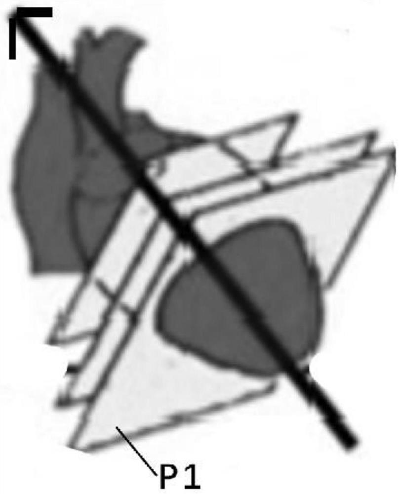
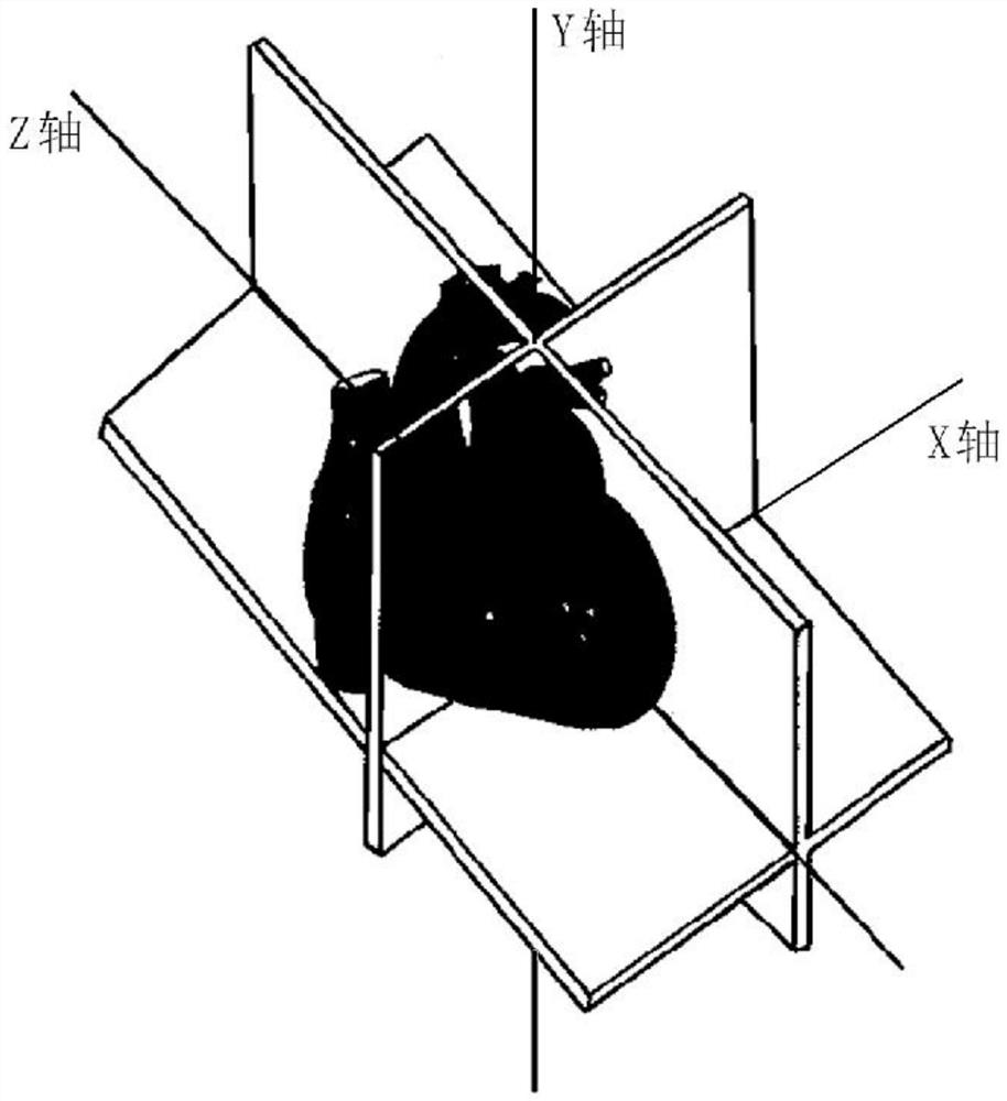
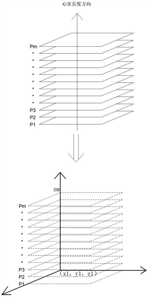
[0044] In the description of the present invention, unless otherwise specified, "plurality" means two or more; unless otherwise specified, "notch-shaped" means a shape other than an even cross-section. The orientation or positional relationship indicated by the terms "upper", "lower", "left", "right", "inner", "outer", "front end", "rear end", "head", "tail" etc. is Based on the orientation or positional relationship shown in the drawings, it is only for the convenience of describing the present invention and simplifying the description, and does not indicate or imply that the referred device or element must have a specific orientation, be constructed and operated in a specific orientation, and th...


PUM


Abstract

The invention discloses a left ventricle center and a region of interest extraction method, a device adopting the method and a system. According to the left ventricle center and region of the interestextraction method, the device adopting the method and the system, the central position of the left ventricle and the stereoscopic region of interest of myocardium are automatically calculated according to an SPECT cross-sectional image, the central position of the left ventricle and the stereoscopic region of interest of the myocardium are enabled to be distinguished from surrounding tissues andartifacts, a judgment result is enabled not to depend on human experience, the result is accurate, the speed is fast, the calculation speed of myocardial SPECT quantitative indexes is increased, manpower is saved, and efficiency is improved.

Description

technical field [0001] The present invention relates to the technical field of SPECT imaging, in particular to a method, device and system for extracting a left ventricular center and a region of interest. Background technique [0002] Emission tomography is a non-invasive nuclear medicine imaging method. Single Photon Emission Computed Tomography (SPECT, Single Photon Emission Computed Tomography) is a type of emission tomography, which has been widely used in pre-clinical drug research and clinical disease diagnosis. . [0003] At present, SPECT / PET is widely used in myocardial tomographic imaging for non-destructive diagnosis of coronary heart disease. However, the traditional qualitative analysis method relies on the naked eye to interpret the image, because it is necessary to analyze dozens of images in the three directions of myocardial short axis X, horizontal long axis Z, and vertical long axis Y, and to compare motion and delay one by one. In this case, the worklo...

Claims


Application Information

Patent Timeline
IPC IPC(8): A61B6/03G06K9/32G06T7/00G06T17/00
CPCA61B6/037A61B6/503A61B6/5211G06T17/00G06T7/0012G06T2207/10108G06T2207/30048G06V10/25
Inventor 杨朝辉王道宇高丽蕾赵文锐郭锋侯岩松黄帅江年铭刘迈
Owner BEIJING NOVEL MEDICAL EQUIP LTD
Why Eureka
Try Eureka