Pathological image processing method and device, storage medium and processor

A technology of pathological image and processing method, applied in the field of image processing, which can solve the problems of low efficiency of pathological image processing and difficulty in obtaining training data

Active Publication Date: 2020-12-01
SHANGHAI XINGMAI INFORMATION TECH CO LTD
View PDF7 Cites 3 Cited by
  • Summary
  • Abstract
  • Description
  • Claims
  • Application Information

AI Technical Summary

Problems solved by technology

[0004] Embodiments of the present invention provide a pathological image processing method, device, storage medium, and processor to at least solve the technical problem of low efficiency in processing pathological images in the prior art due to the difficulty in obtaining model training data

Method used


Image

  • Pathological image processing method and device, storage medium and processor
  • Pathological image processing method and device, storage medium and processor

Examples


Embodiment 1

[0022] According to an embodiment of the present invention, an embodiment of an image processing method is provided. It should be noted that the steps shown in the flowcharts of the accompanying drawings can be executed in a computer system such as a set of computer-executable instructions, and, although A logical order is shown in the flowcharts, but in some cases the steps shown or described may be performed in an order different from that shown or described herein.

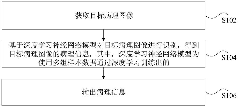
[0023] figure 1 is a flowchart of a pathological image processing method according to an embodiment of the present invention. Such as figure 1 As shown, the method includes the following steps:

[0024] Step S102, acquiring a target pathological image.

[0025] In the technical solution provided in the above step S102 of the present invention, the target pathological image may be an image obtained by photographing a cell sample under a microscope, from which pathological information needs to be identified, w...

Embodiment 2

[0049] The above technical solutions of the embodiments of the present invention will be further illustrated below in combination with preferred implementation manners.

[0050] Deep learning neural networks can be applied to various image processing fields, such as image classification, image segmentation, etc. The deep learning neural network model that meets the needs of practical applications is usually obtained by using a large amount of image data with label information for training based on the initial neural network model.

[0051] In related technologies, some studies have applied deep learning neural network technology to the analysis and processing of cytopathological images, for example, using an image segmentation model to segment cytopathic images to obtain tumor cells. However, to train a good deep learning image segmentation model, a large number of pathological images are required as training data, and professional pathologists are also required to mark the ta...

Embodiment 3

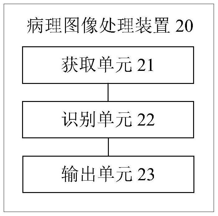
[0059] The embodiment of the present invention also provides an image processing device. It should be noted that the pathological image processing apparatus of this embodiment can be used to implement the pathological image processing method of the embodiment of the present invention.

[0060] figure 2 is a schematic diagram of a pathological image processing device according to an embodiment of the present invention. Such as figure 2 As shown, the pathological image processing device 20 may include: an acquisition unit 21 , an identification unit 22 and an output unit 23 .

[0061] The acquisition unit 21 is configured to acquire a target pathological image, wherein the target pathological image is an image obtained by shooting a cell sample under a microscope.

[0062] The identification unit 22 is used to identify the target pathological image based on the deep learning neural network model to obtain the pathological information of the target pathological image, wherei...


PUM


Abstract

The invention discloses a pathological image processing method and device, a storage medium and a processor. The method comprises the steps that a target pathological image is collected, wherein the target pathological image is an image obtained by shooting a cell sample under a microscope; the target pathological image is recognized based on a deep learning neural network model, and pathologicalinformation of the target pathological image is obtained; wherein the deep learning neural network model is obtained by training a plurality of groups of sample data; wherein each group of data in theplurality of groups of sample data comprises a pathological image sample, a classification label for judging whether the pathological image sample contains malignant cells or not, a segmentation label for judging whether the pathological image sample contains contour information of the malignant cells and a target relationship, and the target relationship is a corresponding relationship among theimage sample, the classification label and the segmentation label; pathological information is output. According to the invention, the technical problem of low pathological image processing efficiency due to the fact that the training data of the model is difficult to obtain in the prior art is solved.

Description

technical field [0001] The present invention relates to the field of image processing, in particular to a pathological image processing method, device, storage medium and processor. Background technique [0002] At present, deep learning neural network technology is applied to the analysis and processing of pathological images. For example, the image segmentation model is used to segment cytopathic images to obtain tumor cells. However, to train a good model, a large number of pathological images are required as training data, and professional pathologists are required to mark the target area of ​​the pathological images used for training, that is, professional pathologists are required to The target region to be segmented is outlined on each pathological image, which is used as training data to train the image segmentation model, which makes it difficult to obtain the training data used for training the model, and thus leads to the technical problem of low efficiency in pro...

Claims


Application Information

Patent Timeline
Patent Type & Authority Applications(China)
IPC IPC(8): G06T7/00G06T7/11G06K9/62
CPCG06T7/0012G06T7/11G06T2207/10056G06T2207/30096G06T2207/20084G06T2207/20081G06F18/24
Inventor 房劬傅琪钲叶德贤
Owner SHANGHAI XINGMAI INFORMATION TECH CO LTD
Why Eureka
Try Eureka