Lesion segmentation method, device and storage medium

A lesion and feature map technology, applied in the field of image recognition, can solve the problems of low lesion segmentation accuracy, affecting the doctor's diagnosis, poor lesion edge segmentation quality, etc. Effect

Active Publication Date: 2021-02-05
PING AN TECH (SHENZHEN) CO LTD
View PDF8 Cites 0 Cited by
  • Summary
  • Abstract
  • Description
  • Claims
  • Application Information

AI Technical Summary

Problems solved by technology

Since the lesion frame can only be segmented with a small feature map size, the segmentation quality of the edge of the lesion is not good, resulting in low accuracy of lesion segmentation, which affects the doctor's diagnosis

Method used


Image

  • Lesion segmentation method, device and storage medium
  • Lesion segmentation method, device and storage medium
  • Lesion segmentation method, device and storage medium

Examples


Embodiment Construction

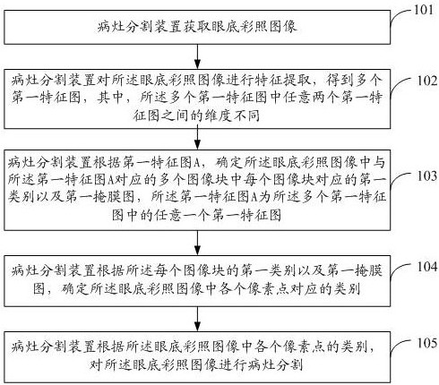
[0029] The following will clearly and completely describe the technical solutions in the embodiments of the present application with reference to the drawings in the embodiments of the present application. Obviously, the described embodiments are part of the embodiments of the present application, not all of them. Based on the embodiments in this application, all other embodiments obtained by persons of ordinary skill in the art without creative efforts fall within the protection scope of this application.

[0030] The terms "first", "second", "third" and "fourth" in the specification and claims of the present application and the drawings are used to distinguish different objects, rather than to describe a specific order . Furthermore, the terms "include" and "have", as well as any variations thereof, are intended to cover a non-exclusive inclusion. For example, a process, method, system, product or device comprising a series of steps or units is not limited to the listed ste...


PUM


Abstract

The present application relates to the technical field of medical science and technology, and specifically discloses a lesion segmentation method, device and storage medium. The method includes: acquiring a fundus color photo image; performing feature extraction on the fundus color photo image to obtain a plurality of first feature maps, wherein the dimensions between any two first feature maps in the plurality of first feature maps are different ; According to the first feature map A, determine the first category and the first mask map corresponding to each image block in the plurality of image blocks corresponding to the first feature map A in the fundus color photo image, the first The feature map A is any first feature map in the plurality of first feature maps; according to the first category and the first mask map of each image block, it is determined that each pixel in the fundus color photo image corresponds to category; according to the category of each pixel in the color fundus photo image, lesion segmentation is performed on the color fundus photo image. The present application is beneficial to improving the accuracy of lesion segmentation.

Description

technical field [0001] The present application relates to the technical field of image recognition, in particular to a lesion segmentation method, device and storage medium. Background technique [0002] Fundus color photo is a way to check the fundus. It can be used to see the organizational structure of the fundus, analyze the normal and abnormal structure of the fundus, and determine whether there are problems with the optic disc, blood vessels, retina or choroid of the fundus. Since image segmentation technology provides rich visual perception information for medical imaging and other applications, it can be applied to the segmentation of lesions related to retinal diseases in fundus color photos. There is a big difference between the lesion segmentation of fundus images and the segmentation of natural images. Due to the influence of shooting light and imaging quality, the contrast of lesion edges is not as clear as that of natural images. Segmentation of fundus lesions ...

Claims


Application Information

Patent Timeline
Patent Type & Authority Patents(China)
IPC IPC(8): G06T7/00G06T7/11G06T7/136G06K9/62G06N3/04G06N3/08
CPCG06T7/0012G06T7/11G06T7/136G06N3/08G06T2207/10024G06T2207/30041G06T2207/20081G06T2207/20084G06N3/047G06F18/2415
Inventor 范栋轶王瑞王立龙王关政
Owner PING AN TECH (SHENZHEN) CO LTD
Why Eureka
Try Eureka