Liver pathological image segmentation model establishment and segmentation method based on attention mechanism

A technology of pathological image and segmentation model, applied in the field of medical image analysis, can solve the problem of inaccurate boundary segmentation between normal tissue area and abnormal tissue area

Active Publication Date: 2020-12-01
NORTHWEST UNIV(CN)
View PDF4 Cites 20 Cited by
  • Summary
  • Abstract
  • Description
  • Claims
  • Application Information

AI Technical Summary

Problems solved by technology

[0005] In order to solve the deficiencies in the prior art, the present invention provides a liver pathological image segmentation model establishment and segmentation method based on the attention mechanism, which solves the problem of under-segmentation in the abnormal and normal tissue areas of the liver histopathological image in the existing segmentation method (Cavities appear) and the problem of inaccurate segmentation of normal tissue areas and abnormal tissue areas

Method used


Image

  • Liver pathological image segmentation model establishment and segmentation method based on attention mechanism
  • Liver pathological image segmentation model establishment and segmentation method based on attention mechanism
  • Liver pathological image segmentation model establishment and segmentation method based on attention mechanism

Examples


Embodiment 1

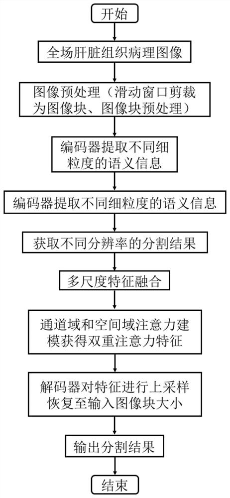
[0084] This embodiment discloses a method for establishing a liver pathological image segmentation model based on an attention mechanism, which specifically includes the following steps:

[0085] Step 1, liver tissue pathological image processing

[0086] The data set in this example is derived from 30 full-field pathological slices of H&E stained liver histopathology collected by the pathology department of a hospital in China. The data set is randomly divided into three parts, and three-fold cross-validation is used to evaluate the network. The folded mean is used as the final result.

[0087] The image processed by the present invention is a digital pathological full-field image (RGB, 3 channels), and the image size is more than 10000╳10000. Due to the limitation of computer performance, it is generally impossible to directly segment the full-field pathological image, so the sliding window strategy is used. Crop the full-field image into 512×512 image blocks. However, in ...

Embodiment 2

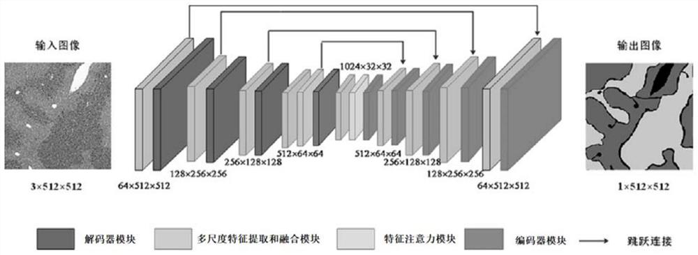
[0124] This embodiment discloses a system for establishing a liver pathological image segmentation model based on an attention mechanism, such as Figure 5 As shown, the system includes:

[0125] (1) The image processing module is used for cropping the liver tissue pathological slice image and its corresponding expert annotated mask image to obtain slice image blocks and mask image blocks; it specifically includes an image cropping module and an image preprocessing module, wherein ,

[0126] (1.1) Image cropping module, which is used for cropping the liver tissue pathological slice image and its corresponding expert annotated mask image,

[0127] (1.2) Image preprocessing module, which is used to uniformly map the background of sliced ​​image blocks and mask image blocks to [255, 255, 255] by setting a threshold, and remove the black area generated by the sliding window strategy clipping to the boundary.

[0128] (2) Segmentation network building module, which is used to con...

Embodiment 3

[0154] This embodiment discloses a liver pathological image segmentation method based on an attention mechanism, which includes the following steps:

[0155] Step 1, the liver pathological image to be processed.

[0156] In this embodiment, 38 groups of 197 groups of liver histopathological image blocks are used as test sets, such as Image 6 The central image shows one of the 38 groups of image blocks. Process according to step 1 in Embodiment 1 to obtain slice image blocks and mask image blocks;

[0157] Step 2, input the obtained slice image block and mask image block obtained in step 1 into the segmentation model obtained in claim 1 to obtain a segmentation probability map, wherein the segmentation probability map includes a target segmentation probability map and a background segmentation probability map; compare The size of the probability value of each pixel on the target segmentation probability map and the background segmentation probability map, and its label is th...


PUM


Abstract

The invention discloses a liver pathological image segmentation model establishment and segmentation method based on an attention mechanism. The method comprises the steps: firstly carrying out the cutting of a liver tissue pathological section image and a corresponding expert labeling mask image, and obtaining a section image block and a mask image block; then constructing a liver tissue pathological image segmentation network based on multi-scale features and an attention mechanism; and taking the slice image blocks and the mask image blocks as inputs of a segmentation network, taking the obtained segmentation probability graph as an output of the segmentation network, and training the obtained segmentation network to obtain a trained segmentation model. And inputting the liver pathological image to be processed into the segmentation model to obtain a segmentation result. According to the segmentation network, a feature attention mechanism is introduced, attention modeling is carriedout on the position and the channel dimension respectively, the distinguishing capacity of the model for a normal tissue area, an abnormal tissue area and a background is improved, and the influenceof many liver tissue pathological image cavities on model learning is relieved.

Description

technical field [0001] The invention belongs to the technical field of medical image analysis, and relates to a method for establishing and dividing a liver pathological image segmentation model based on an attention mechanism. Background technique [0002] Liver injury is a common disease, and in order to treat and study human liver injury, histopathological analysis under the microscope is very important. Due to the large scene, complex background, and numerous tissue areas of pathological slices, manual observation is very time-consuming. At the same time, the analysis of pathological sections is difficult and requires long-term experience accumulation, and the analysis results of pathologists with different experience may be different. With the rapid development of artificial intelligence technology, the use of scanning equipment to digitize pathological slices and automatic analysis with artificial intelligence algorithms has become a research hotspot, mainly focusing ...

Claims


Application Information

Patent Timeline
Patent Type & Authority Applications(China)
IPC IPC(8): G06T7/11G06T7/194G06T7/136G06N3/04G06N3/08
CPCG06T7/11G06T7/194G06T7/136G06N3/08G06T2207/20132G06T2207/20081G06T2207/20084G06T2207/20221G06T2207/30056G06N3/045
Inventor 张墺琦崔磊亢宇鑫武卓越卜起荣
Owner NORTHWEST UNIV(CN)
Why Eureka
Try Eureka