Focus area classification method and system for full-view digital pathological section

A technology for digital pathological slices and region classification, applied in the field of image processing, can solve problems such as low efficiency and time-consuming analysis, and achieve the effects of reducing errors, improving computing efficiency and classification accuracy, and enhancing recognition capabilities

Pending Publication Date: 2020-12-15
XIAMEN UNIV
View PDF3 Cites 12 Cited by
  • Summary
  • Abstract
  • Description
  • Claims
  • Application Information

AI Technical Summary

Benefits of technology

This patented method improves on existing methods by combining two different techniques - one involves identifying specific regions containing abnormalities while another technique uses only those identified region(s). By doing this, models trained from these images have better understanding about how they handle them rather than just focusing their analysis solely upon any single type or location within each pixel's grayscale representation (gray level) alone. Additionally, it suggests setting smaller sizes of pixels around certain parts of an object based on its importance in recognizing important aspects such as disease risk factors. Overall, this approach helps predict diseases faster and accurately without relying heavily on subjective judgment.

Problems solved by technology

The technical problem addressed in this patented text relates how to automatically identify areas where there are abnormalities on histopathology slide sections by analyzing them through image processing techniques like automated microscopy or computers. This can help healthcare professionals make faster diagnoses without having to manually inspect each section individually.

Method used


Image

  • Focus area classification method and system for full-view digital pathological section
  • Focus area classification method and system for full-view digital pathological section
  • Focus area classification method and system for full-view digital pathological section

Examples


Embodiment 1

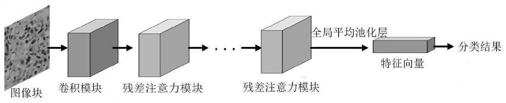
[0027] The invention discloses a method and system for classifying lesion regions of full-field digital pathological slices. The method includes the following steps:

[0028] S1. Annotate the pathological slice image to obtain the annotated slice image;

[0029] Firstly, the acquired original full-view digital pathological slices are cut into smaller pathological slice images; the pathological tissue area in the pathological slice images is segmented by using the Otsu threshold segmentation method, and then the pathological slice images are slid through the pathological slice images using a sliding window of 4096×4096 , and set the threshold to 0.7, filter out the pathological slice images whose pathological tissue area ratio exceeds the threshold, and mark the lesion area.

[0030] S2. The image blocks obtained after twice screening the labeled slice images are used as a training set. In this embodiment, 116 gastric slice images containing gastric cancer regions or common gas...


PUM


Abstract

The invention discloses a lesion area classification method and system for a full-view digital pathological section. The invention builds a CSResNet system, carries out learning training of the CSResNet system, achieves automatic segmentation of a lesion area in the full-view digital pathological section, classify a segmented lesion area, and judges the type of the lesion area. The residual attention module in the CSResNet system is combined with attention mechanisms in the channel direction and the space direction at the same time, so that the network can transfer the learning gravity centerto a key area, capable of deciding the category of an input image, in a feature map, and high calculation efficiency, classification precision and recognition capability are achieved.

Description


Claims


Application Information

Patent Timeline
Owner XIAMEN UNIV
Why Eureka
Try Eureka